

Protein Crystallography from the Perspective of Technology Developments
- PMID: 25983389
- PMCID: PMC4430849
- DOI: 10.1080/0889311X.2014.973868
Protein Crystallography from the Perspective of Technology Developments
Abstract
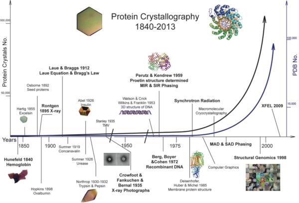
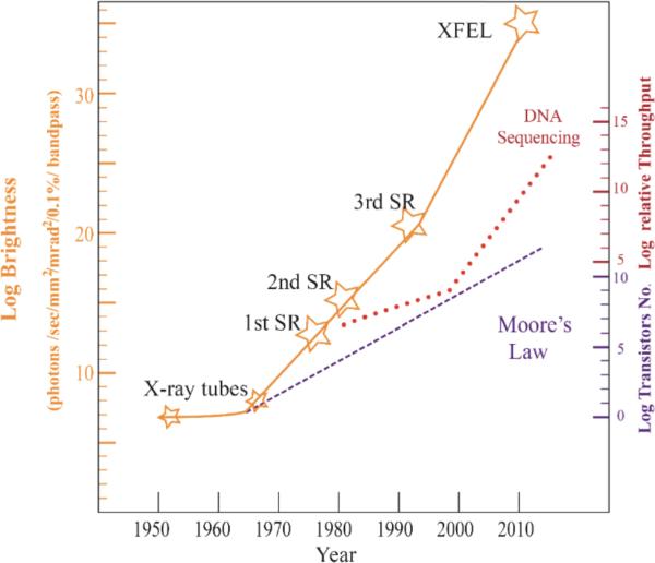
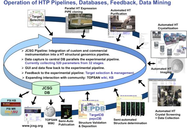
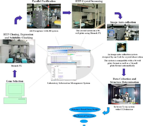
Early on, crystallography was a domain of mineralogy and mathematics and dealt mostly with symmetry properties and imaginary crystal lattices. This changed when Wilhelm Conrad Röntgen discovered X-rays in 1895, and in 1912 Max von Laue and his associates discovered X-ray irradiated salt crystals would produce diffraction patterns that could reveal the internal atomic periodicity of the crystals. In the same year the father-and-son team, Henry and Lawrence Bragg successfully solved the first crystal structure of sodium chloride and the era of modern crystallography began. Protein crystallography (PX) started some 20 years later with the pioneering work of British crystallographers. In the past 50-60 years, the achievements of modern crystallography and particularly those in protein crystallography have been due to breakthroughs in theoretical and technical advancements such as phasing and direct methods; to more powerful X-ray sources such as synchrotron radiation (SR); to more sensitive and efficient X-ray detectors; to ever faster computers and to improvements in software. The exponential development of protein crystallography has been accelerated by the invention and applications of recombinant DNA technology that can yield nearly any protein of interest in large amounts and with relative ease. Novel methods, informatics platforms, and technologies for automation and high-throughput have allowed the development of large-scale, high efficiency macromolecular crystallography efforts in the field of structural genomics (SG). Very recently, the X-ray free-electron laser (XFEL) sources and its applications in protein crystallography have shown great potential for revolutionizing the whole field again in the near future.
Keywords: X-ray crystallography; X-ray free-electron laser (XFEL); computer programs and graphics; protein crystallization; recombinant DNA techniques; structural genomics (SG); synchrotron radiation (SR).
Figures
References
-
- Friedrich W, Knipping P, Laue M. Interferenz-Erscheinungen bei Röntgenstrahlen. Sitz Bayer Akad Wiss. 1912:303–22.
-
- Bragg WH. X-rays and crystals. Nature. 1912;90:219.
-
- Bragg WH, Bragg WL. The reflection of X-rays by crystals. Proc Roy Soc Lond A. 1913;88:428–38.
-
- Bragg WL. The structure of some crystals as indicated by their diffraction of X-rays. Proc R Soc Lond A. 1913;89:248–77.
-
- Thomas JM. William Lawrence Bragg: the pioneer of X-ray crystallography and his pervasive influence. Angewandte Chemie (International ed in English) 2012;51:12946–58. Epub 2012/12/06. - PubMed
Grants and funding
LinkOut - more resources
Full Text Sources
Other Literature Sources
Research Materials
Miscellaneous