

Acute lung injury and the role of histones
- PMID: 25984445
- PMCID: PMC4424229
- DOI: 10.1186/2213-0802-2-1
Acute lung injury and the role of histones
Abstract
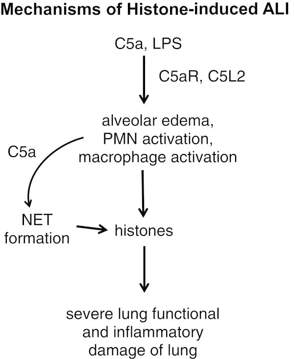
Acute respiratory distress syndrome (ARDS) in humans involves ≥ 200,000 individuals in the United States, and has a mortality rate (40%) for which no specific drug has been approved for use in humans. We have studied experimental acute lung injury (ALI) in mice following airway deposition of bacterial lipopolysaccharide (LPS) or the recombinant mouse complement anaphylatoxin, C5a. As ALI developed over 6 hr, extracellular histones appeared in bronchoalveolar lavage fluids (BALF). Extracellular histone appearance required both C5a receptors (C5aR, C5L2) as well as neutrophils (PMNs) and lung macrophages, as genetic loss of either C5a receptor or depletion of PMNs or macrophages reduced histone levels found in BALF during ALI. It is possible that extracellular histones were derived from formation of neutrophil extracellular traps (NETs) in lung after PMN contact with C5a. When purified histones were delivered to lung via the airways, intense inflammatory injury ensued and type II cells developed large blebs indicating cellular damage and apoptosis. Detailed physiological measurements revealed severe disruption of blood/alveolar gas exchange. These data suggest a key role for histones in development of experimental ALI.
Keywords: ALI; C5a; C5a receptors; Histones.
References
-
- Allam R, Scherbaum CR, Darisipudi MN, Mulay SR, Hagele H, Lichtnekert J, Hagemann JH, Rupanagudi KV, Ryu M, Schwarzenberger C, Hohenstein B, Hugo C, Uhl B, Reichel CA, Krombach F, Monestier M, Liapis H, Moreth K, Schaefer L, Anders HJ. Histones from dying renal cells aggravate kidney injury via TLR2 and TLR4. J Am Soc Nephrol. 2012;23:1375–1388. doi: 10.1681/ASN.2011111077. - DOI - PMC - PubMed
Publication types
Grants and funding
LinkOut - more resources
Full Text Sources
Other Literature Sources
