

Congenital pulmonary airway malformation: A report of two cases
- PMID: 25984523
- PMCID: PMC4419112
- DOI: 10.12998/wjcc.v3.i5.470
Congenital pulmonary airway malformation: A report of two cases
Abstract
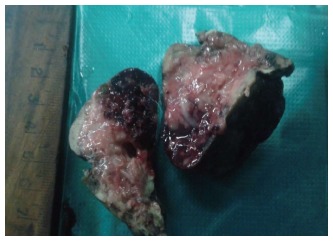
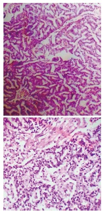
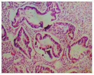
Congenital pulmonary airway malformation (CPAM), previously known as congenital cystic adenomatoid malformation is a congenital disorder of the lung similar to bronchopulmonary sequestration. In CPAM, usually an entire lobe of lung is replaced by a non-working cystic piece of abnormal lung tissue. This abnormal tissue will never function as normal lung tissue. The underlying cause for CPAM is not known. It occurs in approximately 1 in every 30000 pregnancies. The association between CPAM and malignancy has been well documented. There is a small risk (0.7%) of malignant transformation within the cyst. So early diagnosis and surgical resection is important to prevent the grave complications. Herein, we are reporting two interesting cases of CPAM and one belonged to Type II and other belonged to Type III of Stocker's classification.
Keywords: Congenital pulmonary airway malformation-Type II; Congenital pulmonary airway malformation-Type III; Surgical resection.
Figures
References
-
- Stocker JT. The Respiratory Tract. In: Stocker JT, Dehner LP, editors. Pediatric Pathology. 2nd ed. Vol. 1, Chapter 13. Philadelphia: Lippiacott Company; 1992. pp. 466–473.
-
- Sittig SE, Asay GF. Congenital cystic adenomatoid malformation in the newborn: two case studies and review of the literature. Respir Care. 2000;45:1188–1195. - PubMed
-
- Chan C, Lee YS, Taso PC, Jeng MJ, Soong WJ. Congenital Pulmonary Airway Malformation Type IV: A case report. Journal compilation. 2013 Available from: http://www.pedipulm.org.tw/ezcatfiles/rb02/img/img/231/9-2.6.48.pdf.
-
- Sirithangkul S, Chuengchitraks S, Staworn D, Laohapand C, Silarat T. Late manifestation of congenital cystic adenomatoid malformation with lung abscess: a case report. J Med Assoc Thai. 2010;93 Suppl 6:S223–S227. - PubMed
-
- Malik R, Pandya VK, Agarwal G, Jain M. Congenital Cystic AdenomatoidMalformation: A case report. Ind J RadiolImg. 2006;16:859–860.
Publication types
LinkOut - more resources
Full Text Sources
Other Literature Sources
