. 2015;11(5):812-32.
doi: 10.1080/15548627.2015.1034402.

The GST-BHMT assay reveals a distinct mechanism underlying proteasome inhibition-induced macroautophagy in mammalian cells

Affiliations

The GST-BHMT assay reveals a distinct mechanism underlying proteasome inhibition-induced macroautophagy in mammalian cells

Yan-Ning Rui et al. Autophagy. 2015.

Abstract

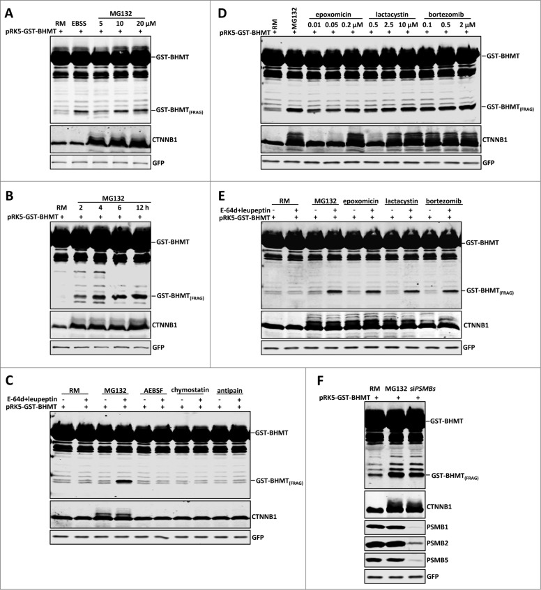
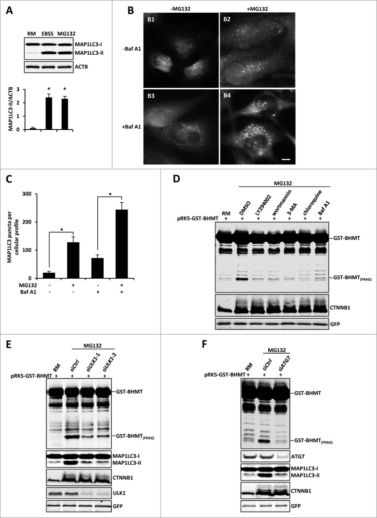
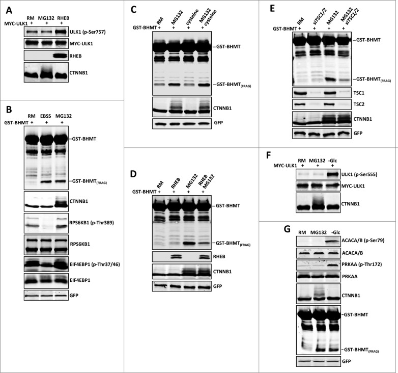
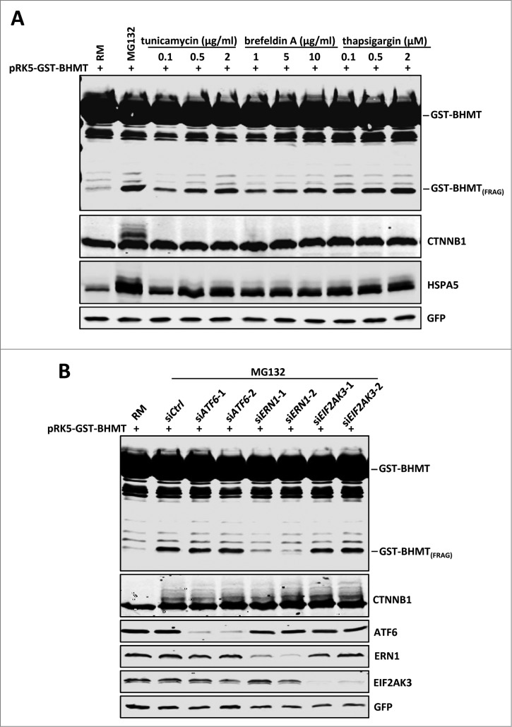
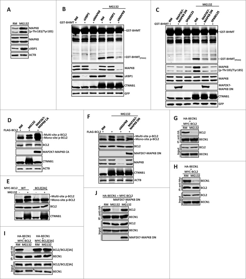
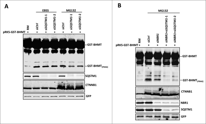
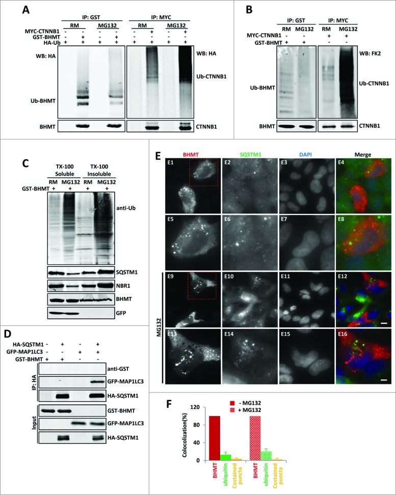
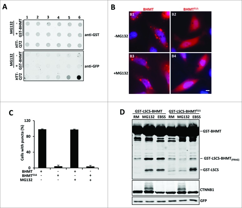
By monitoring the fragmentation of a GST-BHMT (a protein fusion of glutathionine S-transferase N-terminal to betaine-homocysteine S-methyltransferase) reporter in lysosomes, the GST-BHMT assay has previously been established as an endpoint, cargo-based assay for starvation-induced autophagy that is largely nonselective. Here, we demonstrate that under nutrient-rich conditions, proteasome inhibition by either pharmaceutical or genetic manipulations induces similar autophagy-dependent GST-BHMT processing. However, mechanistically this proteasome inhibition-induced autophagy is different from that induced by starvation as it does not rely on regulation by MTOR (mechanistic target of rapamycin [serine/threonine kinase]) and PRKAA/AMPK (protein kinase, AMP-activated, α catalytic subunit), the upstream central sensors of cellular nutrition and energy status, but requires the presence of the cargo receptors SQSTM1/p62 (sequestosome 1) and NBR1 (neighbor of BRCA1 gene 1) that are normally involved in the selective autophagy pathway. Further, it depends on ER (endoplasmic reticulum) stress signaling, in particular ERN1/IRE1 (endoplasmic reticulum to nucleus signaling 1) and its main downstream effector MAPK8/JNK1 (mitogen-activated protein kinase 8), but not XBP1 (X-box binding protein 1), by regulating the phosphorylation-dependent disassociation of BCL2 (B-cell CLL/lymphoma 2) from BECN1 (Beclin 1, autophagy related). Moreover, the multimerization domain of GST-BHMT is required for its processing in response to proteasome inhibition, in contrast to its dispensable role in starvation-induced processing. Together, these findings support a model in which under nutrient-rich conditions, proteasome inactivation induces autophagy-dependent processing of the GST-BHMT reporter through a distinct mechanism that bears notable similarity with the yeast Cvt (cytoplasm-to-vacuole targeting) pathway, and suggest the GST-BHMT reporter might be employed as a convenient assay to study selective macroautophagy in mammalian cells.

Keywords: ACACA/B, acetyl-CoA carboxylase α/β; ACTB, actin, β; ATF4, activating transcription factor 4; ATF6, activating transcription factor 6; ATG7, autophagy-related 7; BCL2, B-cell CLL/lymphoma 2; BECN1, Beclin 1, autophagy-related; BHMT; BHMT, betaine-homocysteine S-methyltransferase; Baf A1, bafilomycin A1; CTNNB1, catenin (cadherin-associated protein), β 1, 88kDa; Cvt, cytoplasm-to-vacuole-targeting; DDIT3, DNA-damage-inducible transcript 3; EBSS, Earle's Balanced Salt Solution; EIF2AK3, eukaryotic translation initiation factor 2-α, kinase 3; EIF4EBP1, eukaryotic translation initiation factor 4E binding protein 1; ER, endoplasmic reticulum; ERN1, endoplasmic reticulum to nucleus signaling 1; GST, glutathionine S-transferase; GST-BHMT(FRAG), an autophagy-mediated cleavage product of the GST-BHMT reporter; GST-BHMT, a fusion protein of glutathionine S-transferase N-terminal to betaine-homocysteine S-methyltransferase; HA, hemagglutinin; HSPA5, heat shock 70kDa protein 5 (glucose-regulated protein, 78kDa); LSCS, linker-specific cleavage site; MAP1LC3, microtubule-associated protein 1 light chain 3; MAP2K7, mitogen-activated protein kinase kinase 7; MAPK8, mitogen-activated protein kinase 8; MTOR; MTOR, mechanistic target of rapamycin (serine/threonine kinase); MTORC1, MTOR complex 1; NBR1, neighbor of BRCA1 gene 1; P4HB, prolyl 4-hydroxylase, β polypeptide; PRKAA, protein kinase, AMP-activated, α catalytic subunit; PRKAA/AMPK; RHEB, Ras homolog enriched in brain; RM, rich medium; RPS6KB1, ribosomal protein S6 kinase, 70kDa, polypeptide 1; SQSTM1, sequestosome 1; TSC1/2, tuberous sclerosis 1/2; ULK1, unc-51 like autophagy activating kinase 1; UPR, unfolded protein response; UPS, ubiquitin proteasome system; XBP1, X-box binding protein 1; cargo receptors SQSTM1/p62 and NBR1; proteasome inhibition; selective macroautophagy.


Figures

Figure 1 (See previous page).
Proteasome inhibition induces GST-BHMT fragmentation. HEK293T cells were transfected with 2 µg pRK5-GST-BHMT plasmids and incubated in the indicated medium followed by coimmunoprecipitation with anti-GST antibody and western blot analyses to detect the proteolytic processing of the GST-BHMT reporter. The prominent accumulation of CTNNB1, an endogenous proteasome substrate, confirmed the effective inactivation of the proteasome by MG132. GFP-MYC, whose expression is driven by the internal ribosome binding sites in the pRK5-GST-BHMT plasmid, was revealed by anti-MYC antibody and served as normalization control, as reported. (A) MG132 treatment induces GST-BHMT fragmentation. HEK293T cells were incubated in EBSS or in nutrient-rich medium (RM) containing MG132 at the indicated concentrations from 5 µM to 20 µM for 6 h, before being processed for the GST-BHMT assay. (B) Time-dependent analysis of GST-BHMT processing. After incubation in nutrient-rich medium with 10 µM MG132, transfected HEK293T cells were harvested at indicated time points from 2 h to 12 h. Notice the gradual accumulation of GST-BHMT(FRAG) product overtime, as revealed by western analysis with anti-GST antibody. (C) Inhibition of lysosomal proteases does not induce GST-BHMT fragmentation. HEK293T cells transfected with GST-BHMT reporter were treated with MG132 (10 µM) or with lysosome cysteine protease inhibitors AEBSF (2 mM), chymostatin (100 µM) or antipain (50 µg/ml), as indicated. Note the apparent accumulation of GST-BHMT(FRAG) only in the sample treated with both MG132 and lysosome protease inhibitors E-64d and leupeptin (lane 4). (D) Multiple proteasome inhibitors, including epoxomicin, lactacystin, and bortezomib, induce similar GST-BHMT processing as that by MG132. (E) The proteasome inhibition-induced accumulation of GST-BHMT(FRAG) requires the presence of lysosomal protease inhibitors E-64d and leupeptin. The transfected cells were treated in the presence or absence of E-64d and leupeptin together with different proteasome inhibitors, as indicated: MG132 (10 µM), epoxomicin (0.1 µM), lactacystin (2.5 µM), and bortezomib (0.5 µM). (F) Genetic interference of proteasomal function leads to similar BHMT fragmentation as that induced by MG132 (compare lanes 3 with 2). For sample in lane 3, cells were cotransfected with 5 nM of siRNAs against proteasome catalytic subunits PSMB1, PSMB2 and PSMB5, and their knockdown efficiency was verified by western blotting analysis, as indicated.
Figure 2 (See previous page).
Proteasome inhibition-induced GST-BHMT processing is autophagy-dependent. (A-C) Proteasome inhibition induces increased levels of MAP1LC3 lipidation and MAP1LC3-positive puncta formation. (A) For MAP1LC3 lipidation assay, HEK293T cells were treated with 10 µM MG132 in the presence of the lysosome inhibitor Baf A1 (100 nM). Both MAP1LC3-I and MAP1LC3-II were enriched by immunoprecipitation using antibody against MAP1LC3. Note that MG132 induced a similar level of MAP1LC3-II as by EBSS treatment. (B-C) For MAP1LC3-positive puncta formation assay, HeLa cells were treated with 10 µM MG132 in the absence or presence of lysosome inhibitor Baf A1 (100 nM), as indicated. Scale bar: 5 µm. (C) Quantitative analysis of cellular MAP1LC3-positive puncta profile. For each sample, MAP1LC3-positive puncta in about 200 cells were counted. The data was presented as the average number of MAP1LC3-positive puncta per cell. (D-F) Proteasome inhibition-induced BHMT processing is autophagy-dependent. (D) HEK293T cells were treated with MG132 (10 µM) together with DMSO control or the following pharmacological inhibitors of the autophagy pathway: LY294002 (100 µM), 3-methyladenine (3-MA; 10 mM), wortmannin (1 µM), chloroquine (100 µM), and Baf A1 (100 nM). Compared to the mock-treated sample using solvent DMSO, all other samples that were simultaneously treated with the indicated autophagy inhibitors showed reduced accumulations of GST-BHMT(FRAG). (E and F) GST-BHMT fragmentation was inhibited following the depletion of essential autophagy components. HEK293T cells were cotransfected with 10 nM siRNA against ULK1 (E) or ATG7 (F). Note that knockdown of either ULK1 (E) or ATG7 (F), but not treatment with control siRNA (siCtrl), significantly reduced the production of GST-BHMT(FRAG) and MAP1LC3 lipidation.
Figure 3.
Figure 3.
Proteasome inhibition-induced GST-BHMT fragmentation is MTOR- and PRKAA-independent. (A to C) Proteasome inhibition does not affect MTORC1 activity. Western blot analysis of the phosphorylation status of MTORC1 substrates (A) ULK1 as well as (B) RPS6KB1 and EIF4EBP1. (A) HEK293T cells were treated with MG132 (10 µM) for 6 h. The phosphorylation status of ULK1 at Ser757 was detected by an antibody specifically against ULK1 (p-Ser757). (B) Compared to the control of untreated cells in nutrient-rich medium (lane 1), MG132 treatment in rich medium did not affect the phosphorylation levels of RPS6KB (p-Thr389) and EIF4EBP1 (p-Thr37/46) (lane 3), whereas EBSS starvation led to significantly reduced levels of phosphorylation at these target sites (lane 2). (C) Cysteine levels do not affect the cellular autophagy activity. In the nutrition-rich medium, addition of cysteine (1 mM) had no effect on the basal level of GST-BHMT fragmentation (compare lane 3 with lane 1) or on MG132-induced GST-BHMT fragmentation (compare lane 4 with lane 2). (D and E) MTOR activation suppresses the proteasomal inhibition-induced autophagy GST-BHMT assays. GST-BHMT (2 µg) was cotransfected with (D) FLAG-RHEB (2 µg) or (E) a mixture of 10 nM siRNAs against TSC1 and TSC2 either in the absence or presence of 10 µM MG132, as indicated. Activation of MTOR, either by (D) overexpression of its activator RHEB or by (E) depletion of its inhibitory TSC1/2 complex, both significantly diminished the MG132-induced fragmentation of the GST-BHMT reporter. The ectopic expression of RHEB and the knockdown efficiency of TSC1 and TSC2 were verified by western blotting against RHEB and TSC1 and TSC2 proteins. (F and G) Proteasome inhibition does not affect PRKAA activity. HEK293T cells treated with MG132 (10 µM) for 6 h were analyzed by western blotting to detect the endogenous phosphorylation levels of (F) ULK1 (p-Ser555), (G) ACACA/B (p-Ser79) and PRKAA (p-Thr172), the known targets of PRKAA. Compared to the controls of untreated samples (lane 1), glucose starvation led to significantly increased phosphorylations of these endogenous PRKAA substrates (lanes 3) whereas MG132 treatment (lane 2) showed little effect, although both treatments similarly resulted in the increased accumulation of GST-BHMT(FRAG).
Figure 4.
Figure 4.
Proteasome inhibition-induced GST-BHMT fragmentation involves the ERN1 branch of the ER stress signaling. (A) ER stress induces GST-BHMT fragmentation. HEK293T cells were treated with MG132 (10 µM) or different ER stress inducers at indicated concentrations, followed by western blotting analyses to examine the proteolytic processing of GST-BHMT reporter and the expression levels of HSPA5, a marker of ER stress. Compared to the untreated control (lane 1), all ER stressors induced robust accumulation of GST-BHMT(FRAG) similar as that by MG132 treatment. (B) ERN1 is critical for the proteasome inhibition-induced BHMT processing. siRNAs against ATF6, ERN1 and EIF2AK3 were transfected into HEK293T cells together with pRK5-GST-BHMT, followed by the GST-BHMT assay. The effectiveness of siRNA-mediated target knockdown was verified by western blotting using the corresponding specific antibodies, as indicated.
Figure 5 (See previous page).
The MAPK8-BCL2-BECN1 pathway is the main mediator downstream of ERN1 in proteasome inhibition-induced autophagy. (A) MG132 activates main downstream effectors of ERN1 signaling: MAPK8 and XBP1. 10 µM MG132 treatment for 2 h induced strong increase of dual-phosphorylated MAPK8 and spliced XBP1 (sXBP1). (B and C) MAPK8 but not XBP1 is required for MG132-induced autophagy. GST-BHMT assays. HEK293T cells were cotransfected with GST-BHMT reporter (2 µg) and (B) siRNAs against XBP1 (30 nM) or MAPK8 (20 nM), or (C) a dominant-negative MAPK8 (MAP2K7-MAPK8 DN, 2 µg) or were treated with SP600125 for 1 h before MG132 treatment. (D and E) MG132 induces multisite phosphorylation of BCL2. (D) Similar to that catalyzed by the constitutively active MAPK8 (MAP2K7-MAPK8 CA, 2 µg), MG132 significantly induced multisite phosphorylation of wild-type BCL2, (E) but not a mutant BCL2 with Thr69Ala and Ser87Ala substitutions. (F) MPAK8 is responsible for the MG132-induced multisite phosphorylation of BCL2. HEK293T cells were treated with 10 µM MG132 for 2 h. Both siRNA-mediated depletion of MAPK8 and expression of MAP2K7-MAPK8 DN significantly reduced the level of multisite phosphorylation in BCL2. (G and H) MG132 treatment induced dissociation between BCL2 and BECN1. HEK293T cells were cotransfected with 2 µg each of MYC-BCL2 and HA-BECN1. Reciprocal coimmunoprecipitation (coIP) assays were performed with (G) anti-HA or (H) anti-MYC antibodies in the absence or the presence of 10 µM MG132 (2 h incubation), as indicated. Note the dramatically reduced interaction between BCL2 and BECN1 in MG132-treated samples. (I and J) MAPK8-mediated multisite phosphorylation of BCL2 is required for the MG132-induced dissociation of BCL2 and BECN1. HA-BECN1 was cotransfected with wild-type MYC-BCL2 or in parallel with (I) mutant MYC-BCL2[3A] (T69A, S70A, S87A), or (J) a dominant-negative MAPK8 (MAP2K7-MAPK8 DN) into HEK293T cells. (I) Contrary to wild-type BCL2, mutant BCL2[3A] remained associated with BECN1 after MG132 treatment. (J) MAP2K7-MAPK8 DN blocked MG132-induced disassociation of BCL2 from BECN1. The knockdown efficiency against target proteins by different siRNAs were verified by western blotting assays. Proteasome inhibition by MG132 was confirmed by the increased accumulation of CTNNB1.
Figure 6.
Figure 6.
Proteasome inhibition-induced GST-BHMT fragmentation requires cargo receptor SQSTM1 and NBR1. (A) SQSTM1 knockdown specifically reduces MG132-induced but not starvation-induced GST-BHMT fragmentation. HEK293T cells were incubated either in EBSS or in nutrient-rich medium (RM) with 10 µM MG132 to induce an autophagic response. (B) NBR1 knockdown reduced MG132-induced GST-BHMT fragmentation (lane 3). Simultaneous knockdown of both NBR1 and SQSTM1 led to a more prominent reduction of GST-BHMT(FRAG) accumulation (lanes 4 and 5). In (A) and (B), 10 nM siRNA against SQSTM1 or NBR1 were cotransfected with the GST-BHMT reporter into the HEK 293T cells and incubated in medium as indicated for 6 h, followed by GST-BHMT assay as described in Figure 1.
Figure 7 (See previous page).
GST-BHMT does not show increased ubiquitination and physical interaction with SQSTM1 in response to proteasome inhibition. (A- C) The ubiquitination patterns of GST-BHMT reporter in response to MG132 treatment. (A and B) HEK293T cells were transfected with pRK5-GST-BHMT reporter (left panels) or the CMV-MYC-CTNNB1 control (right panels), followed by immunoprecipitation with anti-GST or anti-MYC antibodies, as indicated. In (A), cells were cotransfected with CMV-HA-Ub, and the levels of the ubiquitinated GST-BHMT (Ub-BHMT; left panel) or MYC-CTNNB1 (Ub-CTNNB1; right panel) proteins from the cell were detected with anti-HA antibody. In (B), the levels of the ubiquitinated GST-BHMT (Ub-BHMT; left panel) or MYC-CTNNB1 (Ub-CTNNB1; right panel) by endogenous ubiquitin were revealed by FK2 antibody. (C) MG132 does not affect the distribution of GST-BHMT protein in Triton X-100-soluble fractions. The control or MG132-treated HEK293T cells transfected with pRK5-GST-BHMT were harvested into 2 fractions: Triton X-100-soluble (TX-100 soluble) and Triton X-100-insoluble (TX-100 insoluble). The distribution of GST-BHMT, total ubiquitinated proteins, SQSTM1 and NBR1 in both fractions was examined by western blotting using anti-GST, anti-ubiquitin (FK2), anti-SQSTM1 and anti-NBR1 antibodies, respectively, as indicated. The soluble GFP protein, which was hardly detectable in Triton X-100 insoluble fraction, was included as the control for the fractionation procedure. (D) GST-BHMT does not interact with SQSTM1. HEK293T cells were transfected with HA-SQSTM1 together with GST-BHMT or with the positive control GFP-MAP1LC3, followed by immunoprecipitation using anti-HA antibody and western blot detection, as indicated. (E and F) GST-BHMT does not colocalize with SQSTM1. (E) HeLa cells transfected with pRK5-GST-BHMT were immunostained with antibodies against both GST (red) and endogenous SQSTM1 (green) in the absence or presence of MG132, as indicated. Scale bar: 5 µm for E1-E4 and E9-E12; 2 µm for E5-E8 and E13-E16. (F) The quantification of GST-BHMT- and SQSTM1-positive puncta as well as their colocalization profile as described in Materials and Methods.
Figure 8.
Figure 8.
The multimerization domain of GST-BHMT is required for its autolysosomal loading in response to proteasome inhibition. (A) GST-BHMT does not form tight aggregates. Extracts of HEK293T cells transfected with either pRK5-GST-BHMT or CMV-EGFP-Q72-HTT-exon1 were analyzed by filter trap assay using antibodies against GST or GFP, respectively, as indicated. The amount of total proteins loaded in each well was 0, 0.1, 0.4, 2, 20, and 100 µg from lane 1 to lane 6, respectively. While mutant HTT protein formed tight aggregates that were resistant to 6M urea, hardly any such aggregates were detected in extracts from GST-BHMT expressing cells regardless of MG132 treatment. (B and C) Formation of GST-BHMT-positive puncta is dependent on its multimerization domain. (B) HeLa cells transfected with either GST-BHMT or mutant GST-BHMT51Δ were immunostained with anti-GST antibody. Whereas wild-type GST-BHMT protein showed prominent puncta-like subcellular localization, GST-BHMT51Δ was diffusely distributed throughout the cell. Scale bar: 5 µm. (C) Quantification of GST-BHMT-positive puncta, which revealed little change in puncta formation by either wild-type GST-BHMT or mutant GST-BHMT51Δ in response to MG132 treatment. (D) The multimerization domain of GST-BHMT is essential for its autolysosomal loading in response to proteasome inhibition. HEK293T cells were transfected with either pRK5-GST-LSCS-BHMT or mutant pRK5-GST-LSCS-BHMT-Δ51 constructs, followed by EBSS or MG132 treatment and GST-BHMT assay, as indicated.

Similar articles

Cited by

References

    1. Yang Z, Klionsky DJ. Mammalian autophagy: core molecular machinery and signaling regulation. Curr Opin Cell Biol 2010; 22:124–31; PMID:20034776; http://dx.doi.org/10.1016/j.ceb.2009.11.014 - DOI - PMC - PubMed
    1. Ganley IG, Lam du H, Wang J, Ding X, Chen S, Jiang X, et al.. ULK1.ATG13.FIP200 complex mediates mTOR signaling and is essential for autophagy. J Biol Chem 2009; 284:12297–305; PMID:19258318; http://dx.doi.org/10.1074/jbc.M900573200 - DOI - PMC - PubMed
    1. Lamark T, Johansen T. Aggrephagy: selective disposal of protein aggregates by macroautophagy. Int J Cell Biol 2012; 2012:736905; PMID:22518139 - PMC - PubMed
    1. He C, Klionsky DJ. Regulation mechanisms and signaling pathways of autophagy. Annu Rev Genet 2009; 43:67–93; PMID:19653858; http://dx.doi.org/10.1146/annurev-genet-102808-114910 - DOI - PMC - PubMed
    1. Jung CH, Ro SH, Cao J, Otto NM, Kim DH. mTOR regulation of autophagy. FEBS Lett 2010; 584:1287–95; PMID:20083114; http://dx.doi.org/10.1016/j.febslet.2010.01.017 - DOI - PMC - PubMed

Publication types

MeSH terms

Substances