. 2015 Jun;22(6):466-9.
doi: 10.1038/nsmb.3031. Epub 2015 May 18.

The mechanism of inhibition of protein synthesis by the proline-rich peptide oncocin

Affiliations

The mechanism of inhibition of protein synthesis by the proline-rich peptide oncocin

Raktim N Roy et al. Nat Struct Mol Biol. 2015 Jun.

Abstract

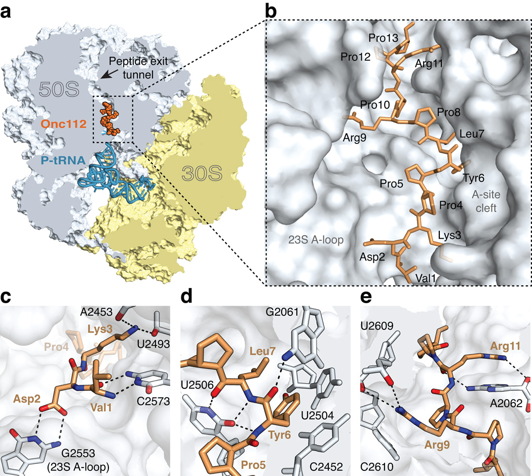
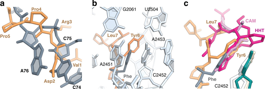
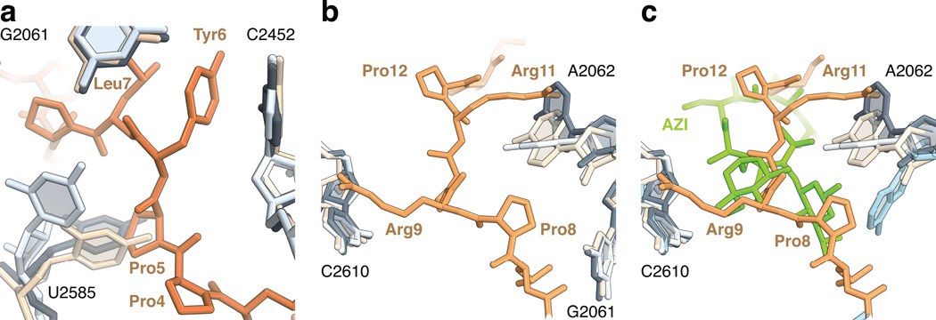
Antibiotic-resistant bacteria are a global health issue necessitating the development of new effective therapeutics. Proline-rich antimicrobial peptides (PrAMPs), which include oncocins, are an extensively studied class of AMPs that counteract bacterial infection at submicromolar concentrations. Oncocins enter and kill bacteria by inhibiting certain targets rather than by acting through membrane lysis. Although they have recently been reported to bind DnaK and the bacterial ribosome, their mode of inhibition has remained elusive. Here we report the crystal structure of the oncocin derivative Onc112 bound to the Thermus thermophilus 70S ribosome. Strikingly, this 19-residue proline-rich peptide manifests the features of several known classes of ribosome inhibitors by simultaneously blocking the peptidyl transferase center and the peptide-exit tunnel of the ribosome. This high-resolution structure thus reveals the mechanism by which oncocins inhibit protein synthesis, providing an opportunity for structure-based design of new-generation therapeutics.


Figures

Figure 1
Figure 1. The structure of onc112 bound to the ribosome
(a) Overview of onc112 (brown) and the P-site tRNAfMet (blue) bound to the 70S ribosome. The 50S and 30S subunits are shown in light blue and yellow, respectively. Portions of the ribosome are omitted for clarity. (b) Close-up view of the positioning of onc112 in the peptide exit tunnel. For reference, landmark features of the ribosome peptidyl transferase center (PTC) are indicated. (c–e) Interactions of onc112 with the ribosome. Putative hydrogen bonds between oxygen (red) and nitrogen (blue) atoms are shown as black dashes. (c) Interactions of the N-terminus of onc112 with the 23S rRNA. (d) Interactions of onc112 within the PTC. The middle part of onc112 occupies the A-site cleft in the PTC. Residues Leu7 and Tyr6 form a three-layer stack with the nucleotide base of C2452 (2463). Additional stabilisation of onc112 is provided by interactions with nucleotides U2506 (2517) and G2061 (2082). (e) Interactions of the C-terminus of onc112 with the peptide exit tunnel. Arg9 and Arg11 form a stacking interaction with the nucleotide bases of C2610 (2621) and A2062 (2083), respectively. PyMOL was used to generate all figures.
Figure 2
Figure 2. Peptide binding to the A-site cleft in the peptidyl transferase center
(a) Residues 1 to 5 of onc112 (brown) follow the path of the CCA-end of an accommodated tRNA in the A site (grey) (PDB accession 1VY4) . (b) The aromatic ring of Tyr6 of onc112 (brown) forms a stacking interaction with the nucleotide base of C2452 (2463) of the 23S rRNA in a manner that is analogous to the phenylalanine residue attached to a Phe-tRNAPhe bound in the A site (grey) (PDB accession 1VY4) . Nucleotides of the 23S rRNA (light blue) forming the A-site cleft are labelled. (c) Structures of antibiotics chloramphenicol (CAM, magenta; 23S rRNA, ivory) (PDB accession 4V7W) and homoharringtonine (HHT, pink; 26S rRNA, darkcyan) (PDB accession 4U4Q) bound in the A-site cleft. Nucleotide C2452 (2463) of the 23S rRNA in the complex of onc112 with the ribosome is light blue.
Figure 3
Figure 3. Conformational changes in the peptide exit tunnel
(a) In the complex of onc112 with the 70S ribosome, the universally conserved nucleotide U2585 (2596) (light blue) changes its position to accommodate Pro5 (70S-CAM complex, ivory, PDB accession 4V7W ; 70S complex with A- and P-site tRNAs, dark grey, PDB accession 1VY4 ). (b) In the upper chamber of the peptide exit tunnel, the binding of onc112 fixes the conformation of the nucleotide base of A2062 (2083) (light blue) such that it forms a stacking interaction with Arg11 and accommodates Pro8. (c) Same as in panel b, but showing the macrolide antibiotic azithromycin (AZI, green) and A2062 (blue) from the structure of the 70S-AZI complex (PDB accession 4V7Y) .

References

    1. Yi HY, Chowdhury M, Huang YD, Yu XQ. Insect antimicrobial peptides and their applications. Appl Microbiol Biotechnol. 2014;98:5807–5822. - PMC - PubMed
    1. de Souza Candido E, et al. The use of versatile plant antimicrobial peptides in agribusiness and human health. Peptides. 2014;55:65–78. - PubMed
    1. Li W, et al. Proline-rich antimicrobial peptides: potential therapeutics against antibiotic-resistant bacteria. Amino Acids. 2014;46:2287–2294. - PubMed
    1. Zasloff M. Antimicrobial peptides of multicellular organisms. Nature. 2002;415:389–395. - PubMed
    1. Brogden KA, Ackermann M, McCray PB, Jr, Tack BF. Antimicrobial peptides in animals and their role in host defences. Int J Antimicrob Agents. 2003;22:465–478. - PubMed

Publication types

MeSH terms

Associated data