. 2015 Aug:60:53-61.
doi: 10.1016/j.biomaterials.2015.04.055. Epub 2015 May 14.

c-Kit+ progenitors generate vascular cells for tissue-engineered grafts through modulation of the Wnt/Klf4 pathway

Affiliations

c-Kit+ progenitors generate vascular cells for tissue-engineered grafts through modulation of the Wnt/Klf4 pathway

Paola Campagnolo et al. Biomaterials. 2015 Aug.

Abstract

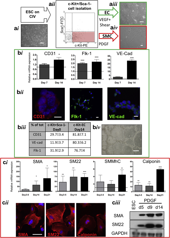
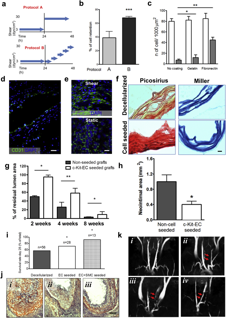
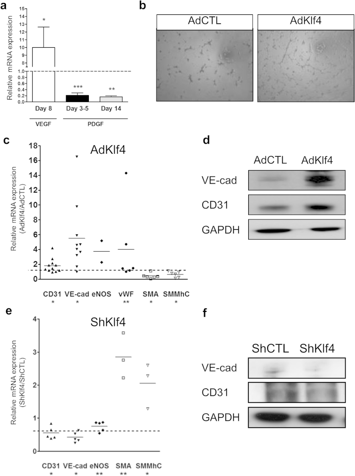
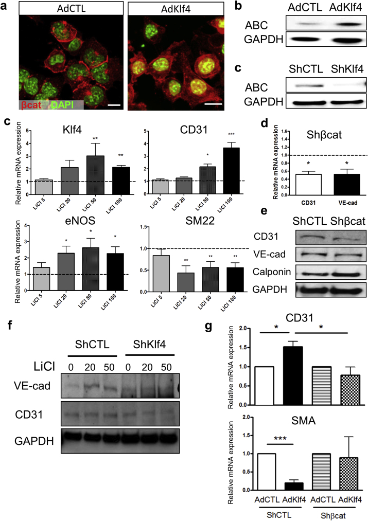
The development of decellularised scaffolds for small diameter vascular grafts is hampered by their limited patency, due to the lack of luminal cell coverage by endothelial cells (EC) and to the low tone of the vessel due to absence of a contractile smooth muscle cells (SMC). In this study, we identify a population of vascular progenitor c-Kit+/Sca-1- cells available in large numbers and derived from immuno-privileged embryonic stem cells (ESCs). We also define an efficient and controlled differentiation protocol yielding fully to differentiated ECs and SMCs in sufficient numbers to allow the repopulation of a tissue engineered vascular graft. When seeded ex vivo on a decellularised vessel, c-Kit+/Sca-1-derived cells recapitulated the native vessel structure and upon in vivo implantation in the mouse, markedly reduced neointima formation and mortality, restoring functional vascularisation. We showed that Krüppel-like transcription factor 4 (Klf4) regulates the choice of differentiation pathway of these cells through β-catenin activation and was itself regulated by the canonical Wnt pathway activator lithium chloride. Our data show that ESC-derived c-Kit+/Sca-1-cells can be differentiated through a Klf4/β-catenin dependent pathway and are a suitable source of vascular progenitors for the creation of superior tissue-engineered vessels from decellularised scaffolds.

Keywords: Cell signalling; Endothelialization; Stem cells; Vascular graft.


Figures

Fig. 1
Fig. 1
Isolation and differentiation of c-Kit+/Sca-1- cells. a: Morphology of differentiated ESC (ai) and flow cytometry strategy for the isolation of c-Kit+/Sca-1- subpopulation (red box, aii). Addition of VEGF and application of shear induced endothelial phenotype (EC, aiii) while PDGF induced smooth muscle cells phenotype (SMC, aiv). b: VEGF treated cells overexpressed EC markers as shown by real time-PCR (bi), immunocytochemistry (bii) and flow cytometry (biii). Functional differentiation was assessed by in vitro tube formation assay (biv). c: PDGF treatment induced expression of SMC markers, as shown at mRNA level (ci) and by immunocytochemistry (cii) and western blot analysis (ciii). Data are expressed as fold-change over Day 0 (dotted lines). Scale bars: 200 μm (a), 25 μm (bii), 100 μm (biv), 50 μm (cii) *P < 0.05; **P < 0.01; ***P < 0.001 vs. Day 0. (For interpretation of the references to colour in this figure legend, the reader is referred to the web version of this article.)
Fig. 2
Fig. 2
Ex vivo seeding of decellularised vessel scaffolds and in vivo grafting. Two protocols were followed to increase the shear from 3 to 30 dyn/cm2 in the tissue engineered grafts after luminal seeding of ECs: ‘daily step-wise’ (protocol A) or ‘hourly step-wise’ (protocol B) (a); cell retention was quantified in (b). Effect of PBS or gelatin or fibronectin coating on cell retention in static (empty bars) and shear (grey bars) was quantified in (c). c-Kit+ ECs formed a homogeneous CD31+ luminal monolayer on the decellularised vessel (d) and elongate in response to shear (e). Picosirius staining for collagen and Miler's elastin staining of decellularised vessels showed improved matrix deposition after cell seeding (f). In vivo implantation of vessels seeded with c-Kit+ ECs showed improved lumen patency (g) and reduced neointima formation (h). Both the seeding with ECs and with the combination of ECs and SMCs (ECs+ SMCs) increased the survival of grafted animals (i) and reduced vessel obstruction (j). MRI detection of the blood flow in the normal carotid (k, i), an occluded carotid grafted with a decellularised vessel (k, ii) and a patent carotid grafted with ECs+ SMCs seeded scaffolds at Day 2 (k, iii), and 2 weeks (k, iv). Red arrows indicate the position of the grafts. Scale bars: 50 μm (d, f, l) and 100 μm (e). *P < 0.05; **P < 0.01; ***P < 0.001. (For interpretation of the references to colour in this figure legend, the reader is referred to the web version of this article.)
Fig. 3
Fig. 3
Effect of Klf4 expression in differentiating c-Kit+ cells. VEGF treatment induced and PDGF decreased Klf4 expression (a, vs. Day 0, dotted line). Overexpression of Klf4 (AdKlf4) increased EC differentiation and reduced SMC markers as shown by tube-formation (b), PCR (c) and western blot (d), as compared to control virus (AdCTL). Klf4 knockdown (ShKlf4) reduced endothelial marker expression and increased levels of SMC markers at RNA (e) and protein level (f) *P < 0.05, **P < 0.001, ***P < 0.0001 vs. control.
Fig. 4
Fig. 4
Klf4 and canonical Wnt pathway are interconnected in the determination of c-Kit+ cells differentiation. Confocal imaging showing the membrane localisation of β-catenin in control cells (AdCTL) and nuclear accumulation in Klf4 overexpressing cells (AdKlf4, a). Western blot confirmed β-catenin activation by AdKlf4 (b) and inhibition by ShKlf4 (c). LiCl (β-catenin activator) induced Klf4 and EC marker expression and reduced the SMC marker expression in a concentration-dependent manner (c: vs. NaCl, dotted line). Knockdown of β-catenin (Shβcat) impaired VEGF-dependent EC differentiation and increased SMC markers (d, vs. control virus: dotted line and e). LiCl-induced increase of EC markers was inhibited by Klf4 knockdown (f, ShKlf4). Klf4-induced EC marker upregulation and SMC marker downregulation is ablated by β-catenin knockdown (g). *P < 0.05; **P < 0.01. Scale bars: 10 μm (a). *P < 0.05; **P < 0.01 and ***P < 0.001.
None
None
None
None
None
None
None

References

    1. Seifu D.G., Purnama A., Mequanint K., Mantovani D. Small-diameter vascular tissue engineering. Nat. Rev. Cardiol. N. D. 2013;10:410–421. - PubMed
    1. Tsai T.-N., Kirton J.P., Campagnolo P., Zhang L., Xiao Q., Zhang Z., Wang W., Hu Y., Xu Q. Contribution of stem cells to neointimal formation of decellularized vessel grafts in a novel mouse model. Am. J. Pathol. 2012;181:362–373. - PubMed
    1. Gong Z., Niklason L.E. Small-diameter human vessel wall engineered from bone marrow-derived mesenchymal stem cells (hMSCs) FASEB J. 2008;22:1635–1648. - PMC - PubMed
    1. Karamariti E., Margariti A., Winkler B., Wang X., Hong X., Baban D., Ragoussis J., Huang Y., Han J.-D.J., Wong M.M., Sag C.M., Shah A.M., Hu Y., Xu Q. Smooth muscle cells differentiated from reprogrammed embryonic lung fibroblasts through DKK3 signaling are potent for tissue engineering of vascular grafts. Circ. Res. 2013;112:1433–1443. - PubMed
    1. Beltrami A.P., Barlucchi L., Torella D., Baker M., Limana F., Chimenti S., Kasahara H., Rota M., Musso E., Urbanek K., Leri A., Kajstura J., Nadal-Ginard B., Anversa P. Adult cardiac stem cells are multipotent and support myocardial regeneration. Cell. 2003;114:763–776. - PubMed

Publication types

MeSH terms