. 2015 Jul;16(7):766-774.
doi: 10.1038/ni.3160. Epub 2015 May 18.

Mechanisms of clonal evolution in childhood acute lymphoblastic leukemia

Affiliations

Mechanisms of clonal evolution in childhood acute lymphoblastic leukemia

Srividya Swaminathan et al. Nat Immunol. 2015 Jul.

Erratum in

Abstract

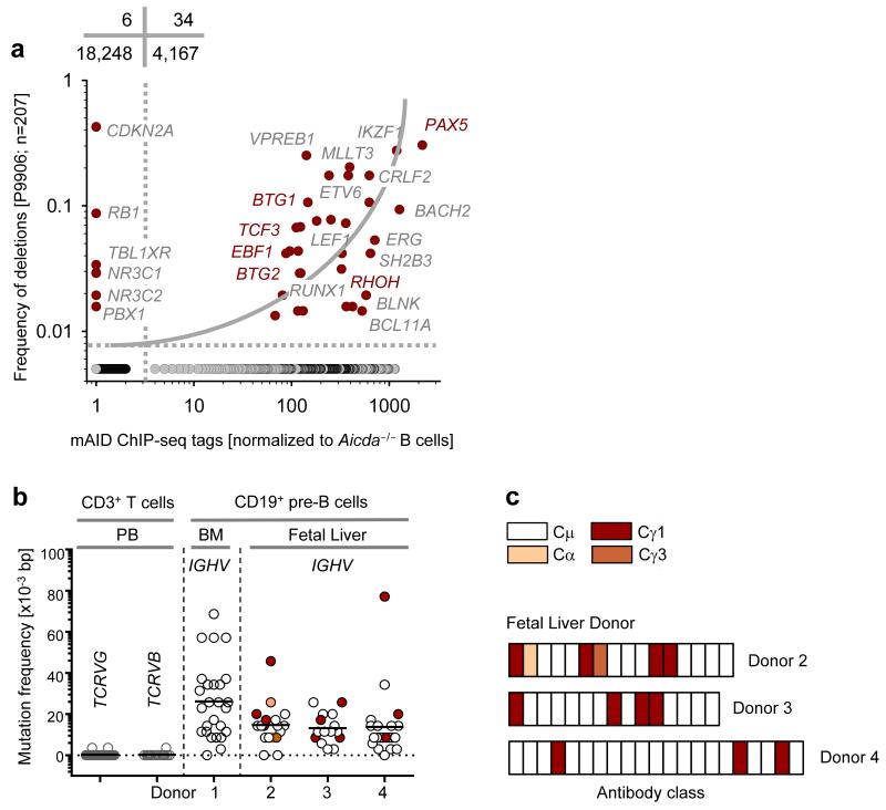
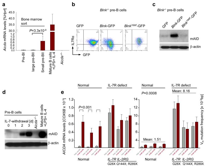
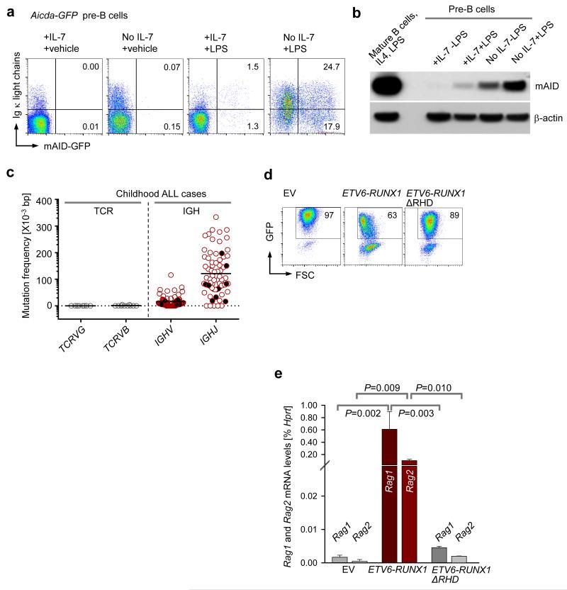
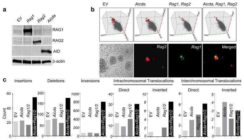
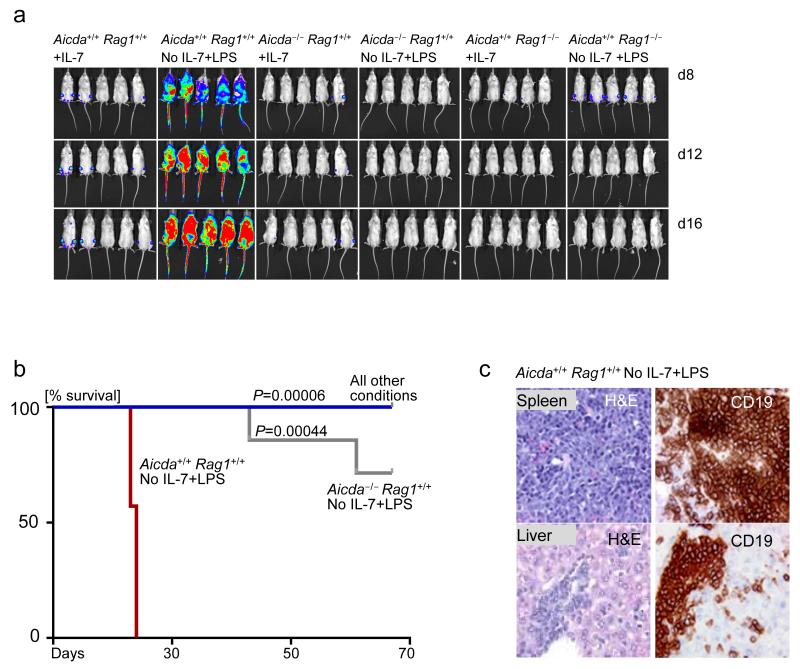
Childhood acute lymphoblastic leukemia (ALL) can often be traced to a pre-leukemic clone carrying a prenatal genetic lesion. Postnatally acquired mutations then drive clonal evolution toward overt leukemia. The enzymes RAG1-RAG2 and AID, which diversify immunoglobulin-encoding genes, are strictly segregated in developing cells during B lymphopoiesis and peripheral mature B cells, respectively. Here we identified small pre-BII cells as a natural subset with increased genetic vulnerability owing to concurrent activation of these enzymes. Consistent with epidemiological findings on childhood ALL etiology, susceptibility to genetic lesions during B lymphopoiesis at the transition from the large pre-BII cell stage to the small pre-BII cell stage was exacerbated by abnormal cytokine signaling and repetitive inflammatory stimuli. We demonstrated that AID and RAG1-RAG2 drove leukemic clonal evolution with repeated exposure to inflammatory stimuli, paralleling chronic infections in childhood.


Figures

Figure 1
Figure 1. Expression and activity of AID in human B cell precursors and B-lineage ALL
(a) Plot showing correlation between common somatic hypermutation targets of mAID and their alteration status in childhood ALL. On plotting the genes which are frequently bound and targeted by mAID, against genes which are commonly deleted and amplified in childhood ALL (data from clinical trial P9906), a positive correlation was observed using a 2×2 contingency table analysis by Chi-square test with Yates’ correction. (b) SHM frequency of IGH VH region genes in single pre-B cell clones isolated from human bone marrow (n=1) and human fetal liver (n=3). SHM of variable regions of TCRVG and TCRVB measured in these cells were used as negative controls. p-values were calculated using student t-test. (c) A schematic depicting CSR in early human B cells derived from three fetal liver donors.
Figure 2
Figure 2. Late pre-B cells (small pre-BII) represent a natural subset of increased genetic vulnerability
(a) Wildtype mouse bone marrow was sorted into different fractions of early B cell development. Quantitative RT-PCR showing Aicda mRNA abundance in each fraction of early B cell development (n=3; mean ± s.d.), as compared to splenic B cells induced with LPS and IL-4 (positive control) and Aicda−/− pre-B cells (negative control). (b) FACS analysis of surface IL-7Rα in Blnk−/− pre-B cells expressing empty GFP vector, Blnk/GFP or BlnkY96F/GFP. (c) Immunoblot depicting increase in mAID protein level upon differentiation of Blnk−/− pre-B cells from large pre-BII to small pre-BII by Blnk reconstitution. (d) mAID protein levels measured by western blotting before (large pre-BII) and after (small pre-BII) IL-7 withdrawal from mouse pre-B cell cultures. (e) Plots showing AICDA mRNA levels (left) and VH mutation frequency (right) in large pre-BII and small pre-BII human pre-B cells, in children with mutation in one of the chains of the IL-7R. Normal human bone marrows were used as negative controls. All p-values were calculated using student t-test. One representative of three experiments is shown for FACS plots and immunoblots.
Figure 3
Figure 3. Aicda and Rag1-Rag2 are regulated by the same transcriptional control elements in pre-B cells
(a) AID protein levels in Stat5fl/fl IL-7-dependent cells transduced with empty ERT2 (no Stat5 deletion) and Cre-ERT2 (with Stat5 deletion) vectors were compared by immunoblotting. (b) Immunoblot for active nuclear FOXO4 after Blnk−/− IL-7-dependent pre-B cells are differentiated from large pre-BII to small pre-BII stage by Blnk reconstitution. (c) Aicda mRNA levels were compared between the empty vector (EV) transduced and Pten deleted Ptenfl/fl pre-B cells 48 hours after tamoxifen induction, by qRT-PCR (n=3, mean ± s.d.). (d-e) Rag1 and Rag2 mRNA levels were measured by qRT-PCR following inducible deletion of Pten in Ptenfl/fl pre-B cells (n=3, mean ± s.d.). (f-h) Mouse IL-7-dependent pre-B cells were retrovirally transduced with either an empty vector or a constitutively active form of Foxo1 (Foxo1CA). The 2 groups of cells were then subject to two conditions each, either they were retained in the presence of IL-7 (large pre-BII) or IL-7 withdrawal was carried out for 24 hours to differentiate them to small pre-BII. (f) Aicda mRNA level was then measured in each case by qRT-PCR (n=3, mean ± s.d.). (g-h) Rag1 and Rag 2 mRNA levels measured by qRT-PCR after retroviral expression of a constitutively active form of Foxo1 (Foxo1CA) or empty vector (EV) in mouse pre-B cells, in the presence or absence of IL-7 (n=3, mean ± s.d.). All p-values were calculated using student t-test.
Figure 4
Figure 4. Evidence for concurrent activities of RAGs and AID in single pre-B cell clones
(a) Aicda-GFP pre-B cells upregulate expression of AID, RAG1 and RAG2 at small pre-BII in the context of inflammatory signals like LPS (GFP+κLC+ cells) (b) Immunoblot comparing mAID protein levels in large pre-BII and small pre-BII mouse pre-B cells, following LPS exposure. Splenocytes from a wildtype mouse induced with LPS and IL-4 were used as a positive control for mAID expression. (c) SHM frequencies of IGH VH regions in pediatric ALL cases were compared against SHM of TCRG and TCRB in these cases. Leukemia patients displaying ongoing VH replacements are represented by solid black circles. (d-e) Effects of ETV6-RUNX1GFP and its mutant ETV6-RUNX1-ΔRHDGFP on Rag1 and Rag2 expression in mouse pre-B cells were evaluated by overexpression of retroviral vectors encoding these fusion proteins. Empty vector (EV) transduced mouse pre-B cells served as negative control. (d) Representative FACS plots showing the percentages of GFP+ cells in each case on day 2 post transduction. One representative of three replicates is shown. (e) GFP+ cells in (d) were sorted and used to measure Rag1 and Rag2 mRNA levels by qRT-PCR (n=3, mean ± s.d.). All p-values were calculated using student t-test.
Figure 5
Figure 5. Cooperation between RAG1, RAG2 and AID promotes clonal evolution towards pre-B ALL
(a) Immunoblot depicting protein expression of AID, RAG1 and RAG2 after retroviral overexpression of these vectors in EBV-immortalized human cord blood B cells. (b) Verification of Aicda (iRFP670), Rag1 (eGFP) and Rag2 (dsRedE2) overexpression in EBV transformed human B cells by flow cytometry (top) and verification of Rag1 and Rag2 overexpression in in these cells by fluorescence microscopy (bottom). (c) Whole exome sequencing analyses to compare the total counts of chromosomal abnormalities in empty vector (EV), Aicda, Rag1-Rag2, and Aicda Rag1-Rag2 transduced EBV-transformed human cord blood B cells.
Figure 6
Figure 6. Cooperation between RAGs and AID is required for clonal evolution of pre-leukemic ETV6-RUNX1 B cell precursors
(a) Luciferase bioimaging of NOD-SCID mice injected with Aicda+/+ Rag1+/+ +IL-7, Aicda+/+ Rag1+/+ No IL-7+LPS, Aicda−/− +IL-7, Aicda−/− No IL-7+LPS, Rag1−/− +IL-7 and Rag1−/− No IL-7+LPS, with all cell types overexpressing ETV6-RUNX1GFP. IL-7 dependent pre-B cells from each group were taken through 5 rounds of IL-7 withdrawal and LPS treatment. 7 mice were used per group. Luciferase bioimages are shown for five mice in each group. (b) Kaplan Meier curves comparing the overall survival percentage of mice in all 6 groups. (c) Verification of pre-B ALL development in the Aicda+/+ Rag1+/+ No IL-7+LPS group by immunohistochemistry. Haematoxylin and Eosin (H&E) staining was carried out to verify lymphocyte infiltration into the liver and spleen of sick mice. Additionally, CD19 immunohistochemistry was carried out to verify that the infiltrating cells represent B-lineage ALL. Immunohistochemistry pictures are shown for one representative mouse out of 7.

References

    1. Wiemels JL, et al. Prenatal origin of acute lymphoblastic leukaemia in children. Lancet. 1999;354:1499–1503. - PubMed
    1. Greaves MF, Wiemels J. Origins of chromosome translocations in childhood leukaemia. Nat. Rev. Cancer. 2003;3:639–649. - PubMed
    1. Bateman CM, et al. Acquisition of genome-wide copy number alterations in monozygotic twins with acute lymphoblastic leukemia. Blood. 2010;115:3553–3558. - PubMed
    1. Greaves M, Maley CC. Clonal evolution in cancer. Nature. 2012;481:306–313. - PMC - PubMed
    1. Gilham C, et al. Day care in infancy and risk of childhood acute lymphoblastic leukaemia: findings from UK case-control study. BMJ. 2005;330:1294. - PMC - PubMed

MeSH terms

Substances