

Accessory spleen in the pelvis: A case report
- PMID: 25985297
- PMCID: PMC4485692
- DOI: 10.1016/j.ijscr.2015.05.009
Accessory spleen in the pelvis: A case report
Abstract
Introduction: Accessory Spleen (AS) is a very rare entity and usually near the spleen's hilum and in the tail of the pancreas. Pelvis reported as an atypical and a rare localization. AS may be formed during embryonic life, they rise from the left side of the dorsal mesogastrium as a result of imperfect fusion of separate splenic masses.
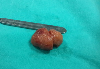
Presentation of case: We report a case of an AS presenting as an left adnexal mass in a middle-aged woman. Transvaginal ultrasonography and magnetic resonance imaging (MRI) revealed a left adnexial mass. Laparatomy was performed, and histological examination revealed that resected mass was splenic tissue.
Discussion: An AS is an incidental finding of no clinical significance in most patients. AS are generally determined during radiological investigations or during open or laparoscopic surgeries. When, the AS settle in the adnexal area; the differential diagnosis could include the causes of adnexal masses like enlarged lymph nodes, subserous fibroid, ovarian tumors, organized hematoma, tuboovarian abscess.
Conclusion: Althought pelvic accessory spleen is a rare condition, should be considered in the differential diagnosis of adnexal masses.
Keywords: Accessory spleen; Adnexal mass; Histopathology.
Copyright © 2015 The Authors. Published by Elsevier Ltd.. All rights reserved.
Figures


References
-
- Unver Dogan N., Uysal I.I., Demirci S., Dogan K.H., Kolcu G. Accessory spleens at autopsy. Clin. Anat. 2011;24(6):757–762. - PubMed
-
- Padilla D., Ramia J.M., Martin J., Pardo R., Cubo T., Hernandez-Calvo J. Acute abdomen due to spontaneous torsion of an accessory spleen. Am. J. Emerg. Med. 1999;17(4):429–430. - PubMed
-
- Radu C.C., Muţiu G., Pop O. Accessory spleen. Rom. J. Morphol. Embryol. 2014;55(3 Suppl):1243–1246. - PubMed
-
- Halpert B., Gyorkey F. Lesions observed in accessory spleens of 311 patients. Am. J. Clin. Pathol. 1959;32(2):165–168. - PubMed
-
- Azar G.B., Awwad J.T., Mufarrij I.K. Accessory spleen presenting as adnexal mass. Acta Obstet. Gynecol. Scand. 1993;72(7):587–588. - PubMed
LinkOut - more resources
Full Text Sources
Other Literature Sources
