. 2015 Jun;21(6):628-37.
doi: 10.1038/nm.3866. Epub 2015 May 18.

KLF4-dependent phenotypic modulation of smooth muscle cells has a key role in atherosclerotic plaque pathogenesis

Affiliations

KLF4-dependent phenotypic modulation of smooth muscle cells has a key role in atherosclerotic plaque pathogenesis

Laura S Shankman et al. Nat Med. 2015 Jun.

Erratum in

Abstract

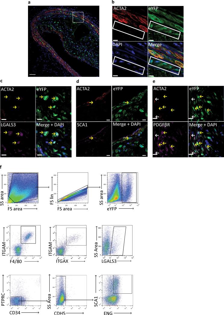
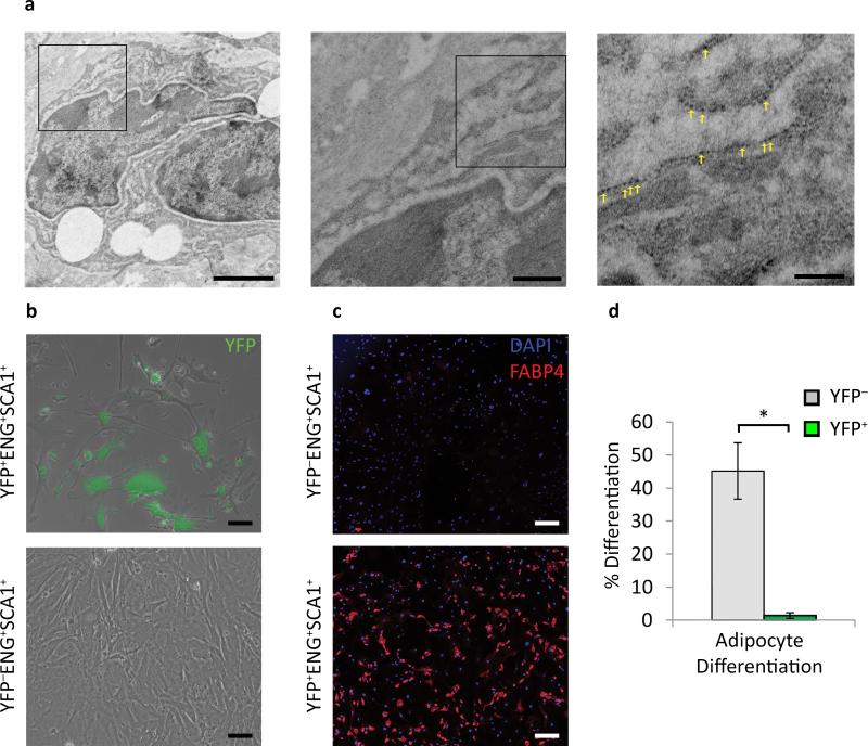
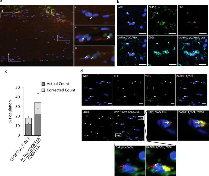
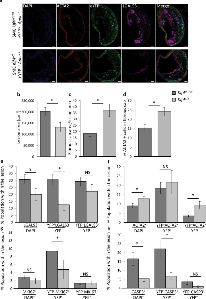
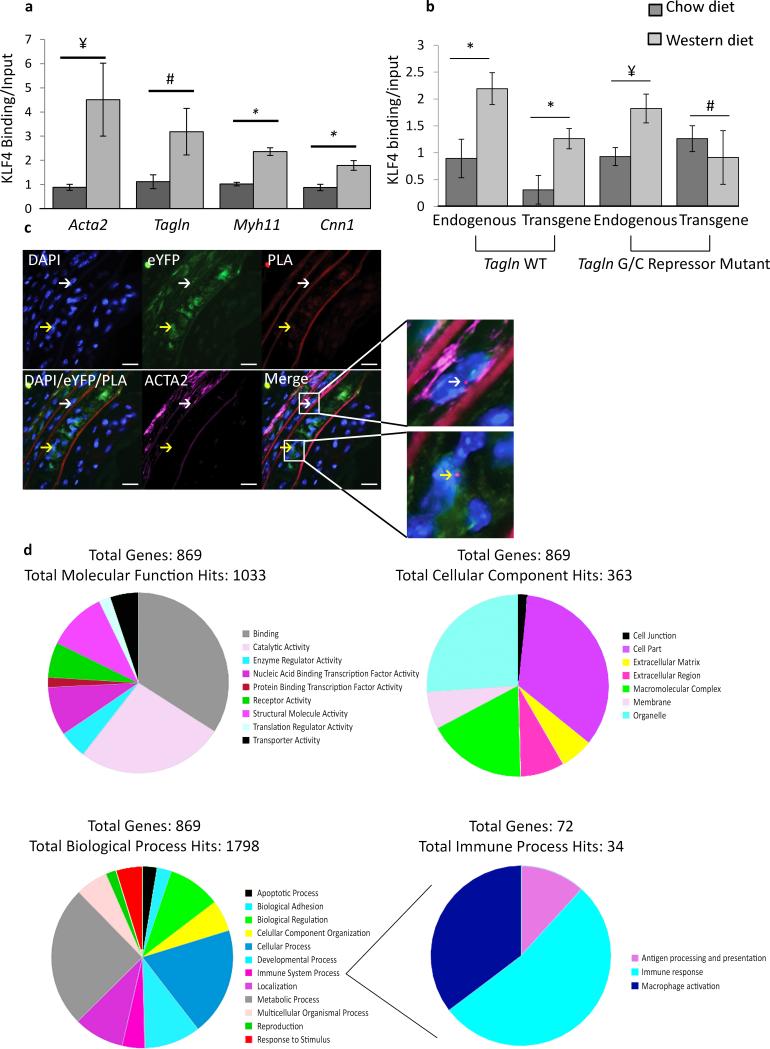
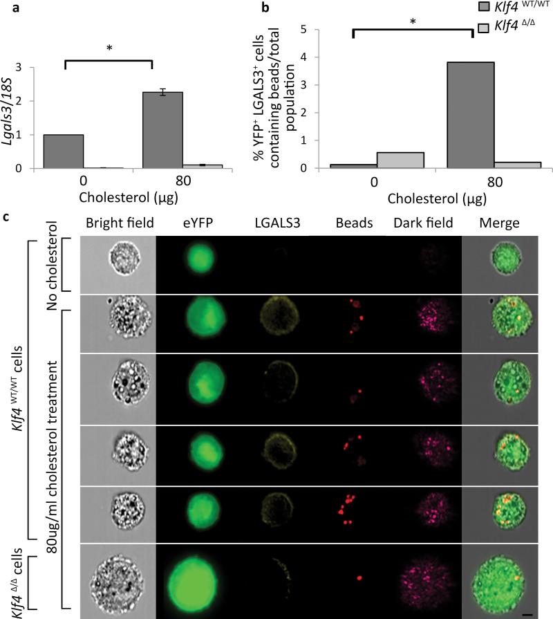
Previous studies investigating the role of smooth muscle cells (SMCs) and macrophages in the pathogenesis of atherosclerosis have provided controversial results owing to the use of unreliable methods for clearly identifying each of these cell types. Here, using Myh11-CreER(T2) ROSA floxed STOP eYFP Apoe(-/-) mice to perform SMC lineage tracing, we find that traditional methods for detecting SMCs based on immunostaining for SMC markers fail to detect >80% of SMC-derived cells within advanced atherosclerotic lesions. These unidentified SMC-derived cells exhibit phenotypes of other cell lineages, including macrophages and mesenchymal stem cells (MSCs). SMC-specific conditional knockout of Krüppel-like factor 4 (Klf4) resulted in reduced numbers of SMC-derived MSC- and macrophage-like cells, a marked reduction in lesion size, and increases in multiple indices of plaque stability, including an increase in fibrous cap thickness as compared to wild-type controls. On the basis of in vivo KLF4 chromatin immunoprecipitation-sequencing (ChIP-seq) analyses and studies of cholesterol-treated cultured SMCs, we identified >800 KLF4 target genes, including many that regulate pro-inflammatory responses of SMCs. Our findings indicate that the contribution of SMCs to atherosclerotic plaques has been greatly underestimated, and that KLF4-dependent transitions in SMC phenotype are critical in lesion pathogenesis.


Figures

Figure 1
Figure 1. SMC lineage tracing provides evidence for large populations of phenotypically modulated SMCs within BCAs of 18 Week WD fed SMC eYFP+/+ Apoe−/− mice
Immunofluorescence staining of representative BCAs from 18 week Western diet fed SMC eYFP+/+ Apoe−/− mice. Results show that differentiated SMCs present at the time of tamoxifen injection (YFP+) subsequently give rise to multiple phenotypes including: (a, b) phenotypically modulated SMCs (YFP+ACTA2 cells highlighted in white rectangle), (c) Mϕ-like SMCs (YFP+ACTA2LGALS3+), (d) mesenchymal stem cell-like SMCs (YFP+ACTA2SCA1+), and (e) MF-like cells (YFP+ACTA2+PDGFβR+). Scale bar represents 50μm (a) and 10μm (b-e). (c-e) Yellow arrows indicate de-differentiated (YFP+/ACTA2) SMCs, white arrows indicate differentiated (YFP+/ACTA2+) SMCs. Samples were either fixed and embedded in paraffin (a-c, e) or Neg40 (d). (f) Flow cytometry of single cell suspension from 18 Week WD fed SMC eYFP+/+ Apoe−/− mouse aortas to further assess phenotypically modulated SMC populations. The gating strategy included: forward versus side scatter gate; a singlets gate; and YFP+ cell gate. Sub-populations of YFP+ cells were found to be double positive for ITGAM (CD11b) and F4/80, ITGAM (CD11b) and ITGAX (CD11c), LGALS3 (mac2), and double positive for SCA1 and ENG after going through lin gating, n = 6 animals.
Figure 2
Figure 2. Phenotypically modulated Mϕ-like SMCs take on some functional characteristics of Mϕs, but MSC-like SMCs show impaired growth and are not multi-potential
(a) Immuno-TEM of BCAs from 18 week Western diet SMC eYFP+/+ Apoe−/− mice using a 10 nm gold bead conjugated secondary antibody revealed lipid laden YFP+ cells engulfing neighboring cells. Yellow arrows indicate immuno-gold beads. Scale bars (from left to right): 2 μm, 0.5 μm, 0.2 μm. (b) Isolated YFP+ and YFP MSCs (SCA1+ENG+) in mesenchymal stem cell media 10 days post plating, green is native YFP signal. (c) YFP+ and YFP MSCs (SCA1+ENG+) after adipogeneisis differentiation based on adipocyte marker FABP4 staining. Scale bar = 200 μm. (d) quantification of adipogenesis from five fields of view per group. Error bars = S.D. * P < 0.05 by Student's t-test.
Figure 3
Figure 3. SMCs within human coronary artery lesions express the Mϕ marker CD68
(a) SMCs within advanced atherosclerotic lesion specimens were identified based on PLA detection of the SMC specific stable epigenetic signature H3K4dime on the MYH11. MYH11 H3K4dime PLA+ cells exhibit a punctate red dot within the nucleus while the non-nuclear amorphous red staining is autofluorescence or non-specific background. (a) Samples were also immuno-stained for CD68 (green), and DAPI (blue). Results showed three distinct cell populations highlighted in enlarged panels to the right and indicated with white arrows: (i) MYH11 H3K4dime PLA+ SMCs that are CD68, (ii) MYH11 H3K4dime PLACD68+ (HSC-derived Mϕs), and (iii) H3K4dime MYH11 H3K4dime PLA+CD68+ SMC-derived Mϕ-like cells. Scale bar = 100 μm. (b) Shoulder regions within plaques [stained with DAPI (blue), ACTA2 (green), PLA (red), and CD68 (cyan)] exhibited a high incidence of SMC-derived Mϕ-like cells (MYH11 H3K4dime PLA+CD68+) (yellow arrows) and several phenotypically modulated SMCs negative for CD68 (MYH11 H3K4dime PLA+ACTA2 CD68) (white arrows). Scale bar = 50 μm. (c) Quantitative analysis of SMC-derived Mϕ-like cells within human coronary lesions based on MYH11 H3K4dime ISH-PLA +/− adjustment for the efficiency of PLA. Error bars = S.E.M. for 12 independent samples of human atherosclerosis in the right coronary artery. (d) Combined epigenetic SMC and genetic HSC lineage tracing analyses of cross gender human heart transplant samples. Coronary artery specimens from a male patient who received a female heart were processed for MYH11 H3K4dime PLA (red), Y-chromosome FISH (green), and CD68 staining (yellow). Results show cells that were MYH11 H3K4dime PLA+ Y-chromosome and CD68+ (yellow arrows) reflecting a SMC-derived Mϕ-like cell not of hematopoietic origin (top). In contrast, Mϕs of hematopoietic origin are MYH11 H3K4dime PLAY-chromosome+CD68+ (red arrows) (bottom). Scale bar = 50 μm.
Figure 4
Figure 4. SMC specific Klf4 conditional KO in Apoe−/− mice fed a high fat diet for 18 weeks resulted in decreased lesion size and increased indices of plaque stability
SMC eYFP+/+ Apoe−/− mice crossed to Klf4FL/FL mice were tamoxifen treated and given Western diet for 18 weeks prior to analysis. (a) Representative image demonstrating the changes in lesion morphology and cell composition. Results showed the following: (b) decreased total lesion area, (c) increased fibrous cap area (defined as the region of the lesion within 30 μm of the luminal surface) relative to the size of the lesion, (d) increased ACTA2+ cells within the fibrous cap, (e) an overall decrease in the fraction of LGALS3+ cells, and a large decrease in the fraction of SMC-derived LGALS3+ cells (YFP+LGALS3+), (f) an increase in the ACTA2+ population within the lesion, (g) decreased proliferation of SMC-derived cells (YFP+MKI67+), and (h) decreased overall cell death, mostly due to a decrease in SMC-derived apoptosis (YFP+CASP3+). *P < 0.05, ¥ P = 0.07, analysis completed by 2-way ANOVA comparing genotyping and distance from start of the BCA with a Tukey post-test, error bars are based on S.E.M. Quantification is based on analysis of five 71415 μm2 regions in each of three sections (d-g), or two sections (h, i), per mouse spanning a 600μm length of the BCA. Scale bar = 50 μm. SMC YFP+/+ Klf4WT/WTApoe−/− n = 11, SMC YFP+/+ Klf4Δ/ΔApoe−/− n = 8.
Figure 5
Figure 5. KLF4 binds to > 800 genes within SMC in advanced atherosclerotic lesions
(a) in vivo ChIP assays on BCAs obtained from 18 week Western diet fed Apoe−/− mice show increased binding of KLF4 to SM marker gene promoters compared to Apoe−/− mice fed a chow diet. P-values based on one-way ANOVA with Tukey post-test. ¥ P = 0.07, # P = 0.11, * P < 0.05. Error bars are based on S.E.M. (b) KLF4 binding to the Tagln promoter in response to Western diet treatment is dependent on the GC repressor element.Apoe−/− mice were crossed to either a Tagln wild type (WT) transgene or Tagln G/C repressor mutant transgene and KLF4 ChIP analyses performed on chromatin extracted from the BCA region. P-values based on two-way ANOVA with Tukey post-test. ¥ P = 0.06, # P = 0.43, * P < 0.05. Error bars are based on S.E.M. n = 3 independent pooled groups of 5 mice per treatment group. (c) KLF4 binding to the G/C repressor element of the Tagln promoter in vivo was determined by Klf4-Tagln ISH-PLA. The white arrow indicates a cell where KLF4 is bound to the Tagln promoter of a differentiated SMC (top), while the yellow arrow identifies a phenotypically modulated SMC (bottom). Scale bar = 10 μm. (d) Aortic segments from the aortic root and aortic arch up to the carotid bifurcations from 18 week WD fed SMC Klf4WT/WT eYFP+/+ Apoe−/− (n = 14), SMC Klf4Δ/Δ eYFP+/+ Apoe−/− (n = 14), and 8 week old chow fed SMC Klf4WT/WT eYFP+/+ Apoe−/− (n = 15) mice were utilized for ChIP-Seq analysis of KLF4 binding targets. 869 targets were enriched in Western diet treated SMC Klf4WT/WT eYFP+/+ Apoe−/− mice as compared to SMC Klf4Δ/Δ eYFP+/+ Apoe−/− mice, and thus represent putative SMC KLF4 target genes.
Figure 6
Figure 6. KLF4-dependent activation of Lgals3, MSC markers, and phagocytotic activity in cholesterol loaded cultured SMC
(a-c) Aortic SMCs were isolated from SMC YFP+/+ Klf4 WT/WT and SMC YFP+/+ Klf4Δ/Δ mice and sorted using a FACSVantage SE DIVA to ensure a pure SMC (YFP+) cell population. (a) Induction of Lgals3 mRNA expression following cholesterol loading (80 μg/mL cholesterol for 72 hours) was decreased in cells derived from SMC YFP+/+ Klf4Δ/Δ as compared to cells derived from SMC YFP+/+ Klf4 WT/WT mice. P-values based on two-way ANOVA with Tukey post-test. *P < 0.05. Data normalized to Klf4 WT/WT 0 ug/mL cholesterol. Error bars based on S.E.M. n = 3 independent experiments. (b-c) Klf4WT/WT and Klf4Δ/Δ cells were incubated with 0.8 μm polysterene beads for 1.5 hours after 72 hours of cholesterol loading to induce a Mϕ-like state. Cells were then analyzed on an Amnis ImagestreamX Mark II to assess expression of YFP, LGALS3, and to identify bead uptake. (b) Quantification of bead uptake was performed using Amnis IDEAS software. After normalization to the total population, values were subjected to Fisher's exact test, which showed that the Klf4WT/WT YFP+LGALS3+ cells contained significantly (P < 0.05) more beads than the Klf4Δ/Δ YFP+LGALS3+ cells (representative experiment from n = 2). (c) Representative images from the Amnis IDEAS software, YFP (green), LGALS3 (yellow), beads (red), Dark Field (magenta). Scale bar = 5 μm.

Similar articles

Cited by

References

    1. Saffitz JE, Schwartz CJ. Coronary atherosclerosis and thrombosis underlying acute myocardial infarction. Cardiol. Clin. 1987;5:21–30. - PubMed
    1. Libby P, Aikawa M. Stabilization of atherosclerotic plaques: new mechanisms and clinical targets. Nat. Med. 2002;8:1257–1262. - PubMed
    1. Falk E, Nakano M, Benton JF, Finn AV, Virmani R. Update on acute coronary syndromes: the pathologists' view. Eur. Heart J. 2012 - PubMed
    1. Falk E, Shah PK, Fuster V. Coronary plaque disruption. Circulation. 1995;92:657–671. - PubMed
    1. Virmani R, Kolodgie FD, Burke AP, Farb A, Schwartz SM. Lessons from sudden coronary death: a comprehensive morphological classification scheme for atherosclerotic lesions. Arterioscler. Thromb. Vasc. Biol. 2000;20:1262–1275. - PubMed

Publication types

Associated data