Review
. 2015 May 18;7(7):a020594.
doi: 10.1101/cshperspect.a020594.

Glia Disease and Repair-Remyelination

Affiliations
Review

Glia Disease and Repair-Remyelination

Robin J M Franklin et al. Cold Spring Harb Perspect Biol. .

Abstract

The inability of the mammalian central nervous system (CNS) to undergo spontaneous regeneration has long been regarded as a central tenet of neurobiology. However, although this is largely true of the neuronal elements of the adult mammalian CNS, save for discrete populations of granular neurons, the same is not true of its glial elements. In particular, the loss of oligodendrocytes, which results in demyelination, triggers a spontaneous and often highly efficient regenerative response, remyelination, in which new oligodendrocytes are generated and myelin sheaths are restored to denuded axons. Yet, remyelination in humans is not without limitation, and a variety of demyelinating conditions are associated with sustained and disabling myelin loss. In this review, we will review the biology of remyelination, including the cells and signals involved; describe when remyelination occurs and when and why it fails and the consequences of its failure; and discuss approaches for therapeutically enhancing remyelination in demyelinating diseases of both children and adults, both by stimulating endogenous oligodendrocyte progenitor cells and by transplanting these cells into demyelinated brain.


Figures

Figure 1.
Figure 1.
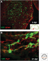
Genetic fate mapping of oligodendrocyte precursor cells (OPCs) reveals them to be the principal source of remyelinating oligodendrocytes. Using Cre-lox fate mapping in transgenic mice, it is possible to show that platelet-derived growth factor receptor α (PDGFRA)/NG2-expressing OPCs (green YFP+) in the adult CNS respond to chemically induced focal demyelination of the ventral spinal cord white matter (inset in A) by proliferation and migration and are abundant within the area of damage, defined here by immunohistochemistry for the astrocyte marker GFAP (red), at 5 d postlesion (dpl) (A). At 21 dpl, when the lesion has undergone complete remyelination, green YFP+ OPC-derived remyelinating oligodendrocytes can be seen producing new myelin sheaths around the demyelinated axons, detected by expression of the myelin protein PLP (red) (B) (see Zawadzka et al. 2010).
Figure 2.
Figure 2.
Systemic delivery of RXR agonists enhance endogenous remyelination in aged animals. Age is a major cause of declining efficiency of endogenous remyelination, largely because of an age-associate decline in the ability of OPCs to differentiate into remyelinating oigodendocytes. The nucleoreceptor RXR-γ is a positive regulator of OPC differentiation. When the RXR agonist 9-cis-retinoic acid is given to aged mice, in which focal demyelination has been generated in the caudal cerebellar peduncles (red outline in brain cross section on left) by injection of ethidium bromide, there is a marked acceleration in the generation of new remyelinating oligodendrocytes compared with saline injected controls. This is evident in the two electron micrographs in which demyelinated axons lack an electron dense myelin sheath, whereas newly remyelinated axons (pseudo-colored in pink) have thin myelin sheaths typical of remyelination that contrast with the thick myelin sheaths of normally myelinated axons evident at the lesion edges (see Huang et al. 2011).
Figure 3.
Figure 3.
Multiple sources of human OPCs can target a broad spectrum of myelin disorders. This schematic shows the major potential sources of myelinogenic central oligodendrocytes and their progenitor cells (OPCs). These include human tissue, embryonic stem cells, induced pluripotential cells, and OPCs directly induced from somatic cells. OPCs may be isolated directly from all of these, on the basis of surface antigens that define several serially generated phenotypes within the oligodendroglial lineage. On transplantation to the brains of afflicted children or adults, the cells may be capable of widespread migration and myelinogenesis. The bottom of the figure provides a list of the major myelin disorders that might be approached through this general strategy of cell-based remyelination from introduced hOPCs. (From Goldman et al. 2012; adapted, with permission, from the author.)
Figure 4.
Figure 4.
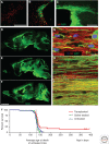
Fetal- and adult-derived human OPCs are distinct in their remyelination competence. (A) Fetal-derived human hNA+ OPCs (red) expressed no detectable MBP at 4 wk after neonatal graft into myelin-deficient and immunodeficient shiverer (MBPshi/shi) × rag2−/− mice; no myelin development was noted in these mice until 12 wk. (B) In contrast, adult-derived OPCs (red) achieved dense MBP expression (green) by 4 wk after neonatal graft. (C) Low-power coronal images of callosal–fimbrial junction of shiverer recipient, showing dense myelination 12 wk after perinatal graft of adult human OPCs. (D–F) Sagittal sections of fetal hOPC-implanted mice immunolabeled for MBP (green) at (D) 20 wk, (E) 35 wk, and (F) 52 wk. Fetal hOPCs dispersed more effectively and expanded more than adult OPCs. By 1 yr, fetal hOPC-derived myelination appeared complete throughout the forebrain and hindbrain, and indeed the entire central neuraxis. (GI) Corresponding confocal optical sections of transplanted shiverer mouse corpus callosum taken at (G) 20 wk, (H) 35 wk, and (I) 52 wk after fetal hOPC graft immunolabeled for neurofilament (red) and MBP (green), reveal the progressive increase in axonal ensheathment with time. (J) Kaplan–Meier survival plot of immunodeficient shiverer mice, either engrafted with human OPCs at birth (red), injected with saline (green), or untreated (blue). Most died between 18 and 21 wk. However, a fraction of engrafted mice (23% to >1 yr in this series) lived substantially longer than any control mouse; these rescued mice have lived normal life spans (typically >2 yr), with substantial recovery of neurological phenotypic. Scale bars, 100 μm (A); 1 mm (C); 2.5 mm (F); 10 μm (G). (Images AC from Windrem et al. 2004 and DI from Windrem et al. 2008; reprinted, with permission, from the authors.)
Figure 5.
Figure 5.
hiPSC OPCs are efficient agents for therapeutic myelination. High-power confocal images of the callosal white matter of mice engrafted with hiPSC OPCs. (A) Dense donor-derived myelination of single axons is evident, with myelin basic protein (MBP, green) production by donor-derived cells (human nuclear antigen, red), and (B) ensheathment of host axons (neurofilament, red) by donor-derived myelin (green). (C) iPSC OPC-derived oligodendrocytic differentiation and myelination permitted the composition of architecturally appropriate nodes of Ranvier in transplanted shiverers. Oligodendrocytic paranodal Caspr protein (red) is seen here flanking βIV spectrin (green)-defined nodes. (D,E) Electron micrographs of iPSC oligodendrocyte-derived myelin in the corpus callosum at 40 wk. Thick myelin wraps with alternating major dense and intraperiod lines, characteristic of mature myelin, are evident. (F,G) hiPSC OPC dispersal and donor-derived myelination of shiverer forebrain. (F) Dot map indicating the distribution of hiPSC OPC-derived cells at 7 mo, following neonatal engraftment. Widespread dispersal and chimerization by hiPSC OPCs is evident (hNA, red). (G) Extensive donor-derived (MBP, green) myelination is evident; sampled 1 mm lateral to F. MBP immunoreactivity (green) is all donor derived. (H) Kaplan–Meier plot of the survival of iPSC-OPC-implanted versus saline-injected mice. Remaining engrafted mice were killed for electron microscopy at ≥270 d. Scale bars, A and B, 5 µm; C, 5 µm; D, 200 nm; E, 100 nm; F and G, 2 mm.
Figure 6.
Figure 6.
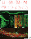
CD140-isolated OPCs are able to broadly migrate and efficiently myelinate. (A) Computer-assisted drawings of 14-μm sections of a 12-wk-old shiverer × rag2-null mouse, transplanted bilaterally in the corpus callosum with 100,000 CD140a+ cells. Red dots represent individual cells labeled with antihuman nuclear antigen. (B) The corpus callosum of an engrafted shiverer mouse at 12 wk, stained for MBP, showing substantial donor-derived myelin. (C) A photomicrograph of the corpus callosum and fimbria in another engrafted mouse. (D) An individual oligodendrocyte, stained for antihuman nuclear antigen (red). (E,F) Ensheathment of host mouse axons (neurofilament, green) at 12 wk by CD140a+ (E) or A2B5+ (F) human fetal cells, showing the more rapid and robust axonal myelination by CD140a+ cells. (G) A CD140a+ cell-engrafted shiverer callosum at 12 wk, immunostained for MBP, human GFAP, and human nuclear antigen, showing robust production of hGFAP+ astrocytes as well as MBP+ oligodendrocytes. Scale bars, B, 500 µm; C, 200 µm; D, 10 µm; E and F, 20 µm. (Images from Sim et al. 2011; reprinted, with permission, from the authors.)

References

    1. Albert M, Antel J, Bruck W, Stadelmann C. 2007. Extensive cortical remyelination in patients with chronic multiple sclerosis. Brain Pathol 17: 129–138. - PMC - PubMed
    1. Altucci L, Leibowitz MD, Ogilvie KM, de Lera AR, Gronemeyer H. 2007. RAR and RXR modulation in cancer and metabolic disease. Nat Rev Drug Discov 6: 793–810. - PubMed
    1. Armstrong RC, Le TQ, Frost EE, Borke RC, Vana AC. 2002. Absence of fibroblast growth factor 2 promotes oligodendroglial repopulation of demyelinated white matter. J Neurosci 22: 8574–8585. - PMC - PubMed
    1. Armstrong RC, Le TQ, Flint NC, Vana AC, Zhou YX. 2006. Endogenous cell repair of chronic demyelination. J Neuropathol Exp Neurol 65: 245–256. - PMC - PubMed
    1. Arnett HA, Mason J, Marino M, Suzuki K, Matsushima GK, Ting JPY. 2001. TNFα promotes proliferation of oligodendrocyte progenitors and remyelination. Nat Neurosci 4: 1116–1122. - PubMed

Publication types

LinkOut - more resources