. 2015 Aug 1;197(15):2489-98.
doi: 10.1128/JB.00268-15. Epub 2015 May 18.

Biochemical Characterization of Mycobacterium smegmatis RnhC (MSMEG_4305), a Bifunctional Enzyme Composed of Autonomous N-Terminal Type I RNase H and C-Terminal Acid Phosphatase Domains

Affiliations

Biochemical Characterization of Mycobacterium smegmatis RnhC (MSMEG_4305), a Bifunctional Enzyme Composed of Autonomous N-Terminal Type I RNase H and C-Terminal Acid Phosphatase Domains

Agata Jacewicz et al. J Bacteriol. .

Abstract

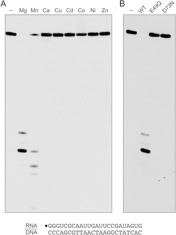
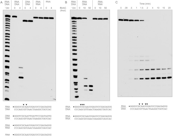
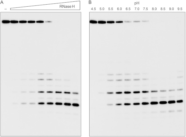
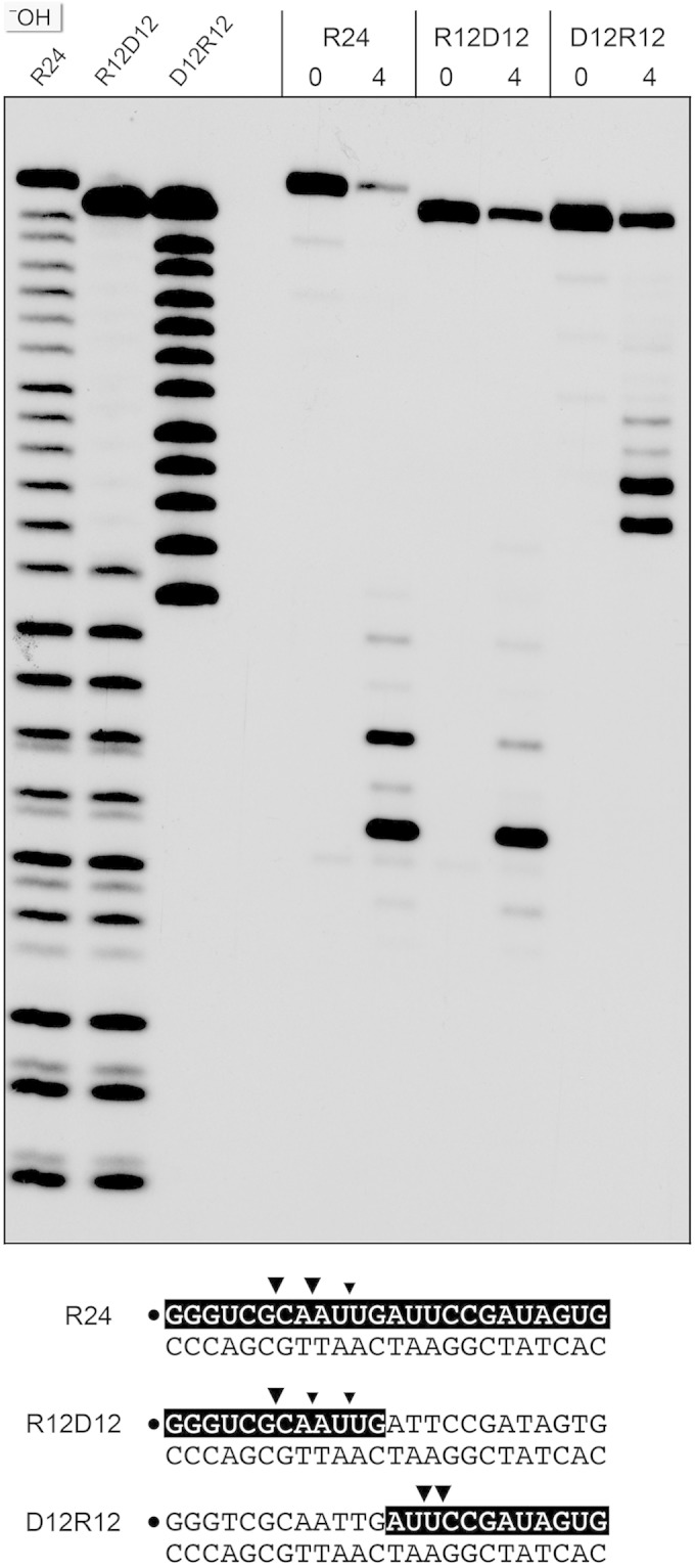
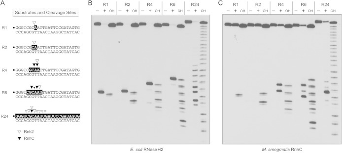
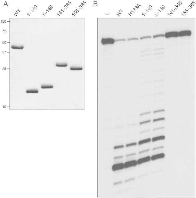
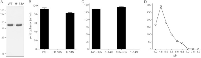
Mycobacterium smegmatis encodes several DNA repair polymerases that are adept at incorporating ribonucleotides, which raises questions about how ribonucleotides in DNA are sensed and removed. RNase H enzymes, of which M. smegmatis encodes four, are strong candidates for a surveillance role. Here, we interrogate the biochemical activity and nucleic acid substrate specificity of M. smegmatis RnhC, a bifunctional RNase H and acid phosphatase. We report that (i) the RnhC nuclease is stringently specific for RNA:DNA hybrid duplexes; (ii) RnhC does not selectively recognize and cleave DNA-RNA or RNA-DNA junctions in duplex nucleic acid; (iii) RnhC cannot incise an embedded monoribonucleotide or diribonucleotide in duplex DNA; (iv) RnhC can incise tracts of 4 or more ribonucleotides embedded in duplex DNA, leaving two or more residual ribonucleotides at the cleaved 3'-OH end and at least one or two ribonucleotides on the 5'-PO4 end; (v) the RNase H activity is inherent in an autonomous 140-amino-acid (aa) N-terminal domain of RnhC; and (vi) the C-terminal 211-aa domain of RnhC is an autonomous acid phosphatase. The cleavage specificity of RnhC is clearly distinct from that of Escherichia coli RNase H2, which selectively incises at an RNA-DNA junction. Thus, we classify RnhC as a type I RNase H. The properties of RnhC are consistent with a role in Okazaki fragment RNA primer removal or in surveillance of oligoribonucleotide tracts embedded in DNA but not in excision repair of single misincorporated ribonucleotides.

Importance: RNase H enzymes help cleanse the genome of ribonucleotides that are present either as ribotracts (e.g., RNA primers) or as single ribonucleotides embedded in duplex DNA. Mycobacterium smegmatis encodes four RNase H proteins, including RnhC, which is characterized in this study. The nucleic acid substrate and cleavage site specificities of RnhC are consistent with a role in initiating the removal of ribotracts but not in single-ribonucleotide surveillance. RnhC has a C-terminal acid phosphatase domain that is functionally autonomous of its N-terminal RNase H catalytic domain. RnhC homologs are prevalent in Actinobacteria.


Figures

FIG 1
FIG 1
Recombinant RnhC. (A) Primary structure. The amino acid sequence of M. smegmatis (Msm) MSMEG_4305/RnhC is aligned with that of M. tuberculosis (Mtu) Rv2228c. The positions of side chain identity are denoted by dots above the residues. Gaps in the alignments are denoted by dashes. The Glu49 and Asp73 RNase H active-site residues and the His173 phosphatase active-site residue that were mutated are shaded in black. The margins of the autonomous N-terminal RNase H and C-terminal acid phosphatase domains, as defined by deletion analysis, are indicated by reverse and forward arrows above the RnhC sequence. (B) Purification. Aliquots (4 μg) of recombinant wild-type RnhC (WT) and E49Q and D73N mutants were analyzed by SDS-PAGE. The Coomassie blue-stained gel is shown. The positions and sizes (in kilodaltons) of marker polypeptides are indicated on the left.
FIG 2
FIG 2
Metal-dependent RNase H activity. (A) Reaction mixtures (10 μl) containing 50 mM Tris-HCl, pH 7.5, 50 mM NaCl, 20 nM (200 fmol) 32P-RNA:DNA hybrid duplex (depicted at the bottom), 0.4 nM (4 fmol) wild-type RnhC, and 5 mM the indicated divalent cation (as the chloride salt) were incubated at 37°C for 20 min. Divalent cation was omitted from a control reaction in lane −. (B) Reaction mixtures (10 μl) containing 50 mM Tris-HCl (pH 7.5); 50 mM NaCl; 10 mM MgCl2; 1 mM DTT; 20 nM (200 fmol) RNA:DNA hybrid duplex; and 0.4 nM (4 fmol) wild-type RnhC, RnhC-E49Q, or RnhC-D73N were incubated at 37°C for 20 min. RnhC was omitted from a control reaction in lane −. The reactions were quenched with an equal volume of 90% formamide, 50 mM EDTA, 0.3% bromophenol blue. The reaction products were analyzed by electrophoresis through a 40-cm 18% polyacrylamide gel containing 7 M urea in 45 mM Tris-borate, 1 mM EDTA. The products were visualized by autoradiography.
FIG 3
FIG 3
Substrate specificity and kinetics. (A and B) Reaction mixtures (10 μl) containing 25 mM Tris-HCl (pH 7.5); 50 mM NaCl; either 10 mM MgCl2 (A) or 10 mM MnCl2 (B); 1 mM DTT; either 200 fmol 32P-RNA:DNA, 32P-DNA:DNA, or 32P-RNA:RNA duplexes or 200 fmol 32P-RNA single strand (as depicted at the bottom, with the 5′ radiolabel denoted by a dot); and RnhC as specified were incubated at 37°C for 20 min. The products were analyzed by urea-PAGE and visualized by autoradiography. An alkaline hydrolysis ladder of the 32P-labeled 24-mer RNA strand was analyzed in parallel in lanes OH. (C) Time course of RNA:DNA cleavage. Reaction mixtures (110 μl) containing 50 mM Tris-HCl (pH 7.5), 50 mM NaCl, 10 mM MgCl2, 1 mM DTT, 20 nM 32P-RNA:DNA hybrid duplex (depicted at the bottom), and 0.4 nM RnhC were incubated at 37°C. Aliquots (10 μl) were withdrawn at the times specified and quenched with formamide-EDTA. The reaction products were analyzed by urea-PAGE and visualized by autoradiography. The principal sites of RNase H incision are indicated by arrowheads above the 32P-labeled RNA strands shown at the bottom.
FIG 4
FIG 4
Enzyme dependence and effect of pH. (A) RnhC titration. Reaction mixtures (10 μl) containing 50 mM Tris-HCl (pH 7.5), 50 mM NaCl, 10 mM MgCl2, 1 mM DTT, 20 nM (200 fmol) 32P-RNA:DNA hybrid duplex, and increasing amounts of RnhC (0.02, 0.04, 0.1, 0.2, 0.4, 1, 2, and 4 fmol, from left to right) were incubated at 37°C for 20 min. RnhC was omitted from a control reaction in lane −. (B) pH profile. Reaction mixtures (10 μl) containing 50 mM buffer {either Tris-acetate, pH 4.5, 5.0, 5.5, 6.0, or 6.5; Tris-HCl, pH 7.0, 7.5, 8.0, 8.5, or 9.0; or CHES [2-(cyclohexylamino)ethanesulfonic acid], pH 9.5}, 50 mM NaCl, 10 mM MgCl2, 1 mM DTT, 20 nM (200 fmol) 32P-RNA:DNA duplex, and 0.4 nM (4 fmol) RnhC were incubated at 37°C for 5 min. The reaction products were analyzed by urea-PAGE and visualized by autoradiography.
FIG 5
FIG 5
Cleavage of chimeric RNA-DNA junction substrates. Reaction mixtures (10 μl) containing 50 mM Tris-HCl (pH 8.0); 50 mM NaCl; 10 mM MgCl2; 1 mM DTT; 200 fmol 32P-labeled 24-mer duplex R24, R12D12, or D12R12 (shown at the bottom, with the 32P label denoted by a dot and the ribonucleotides shaded black); and 0 or 4 fmol RnhC was incubated for 20 min at 37°C. The products were resolved by urea-PAGE and visualized by autoradiography. Alkaline hydrolysis ladders of the 32P-labeled R24, R12D12, and D12R12 strands were analyzed in parallel in the three lanes on the left (OH). Major and minor cleavage sites are denoted by large and small arrowheads, respectively.
FIG 6
FIG 6
Minimum RNA requirement. (A) Substrates and cleavage sites. The 32P label is denoted by a dot. Ribonucleotides are shaded in black. Sites of incision by E. coli RNase H2 and M. smegmatis RnhC are indicated by open and solid arrowheads, respectively. (B) Reaction mixtures (10 μl) containing 20 mM Tris-HCl (pH 8.8), 10 mM (NH4)2SO4, 10 mM KCl, 2 mM MgSO4, 0.1% Triton X-100, 20 nM (200 fmol) duplex substrate as specified and either no enzyme (lanes −) or 5 U E. coli RNase H2 (lanes +) were incubated at 37°C for 20 min. (C) Reaction mixtures (10 μl) containing 50 mM Tris-HCl (pH 8.0), 50 mM NaCl, 10 mM MgCl2, 1 mM DTT, 20 nM (200 fmol) duplex substrate as specified, and either no enzyme (lanes −) or 5 nM (50 fmol) RnhC were incubated at 37°C for 20 min. The products were analyzed by urea-PAGE and visualized by autoradiography. Alkaline hydrolysis ladders of the 32P-labeled strands were analyzed in parallel in lanes OH.
FIG 7
FIG 7
RNase H activities of the isolated domains. (A) Aliquots (5 μg) of full-length RnhC (WT), RnhC-(1-140), RnhC-(1-149), RnhC-(141-365), and RnhC-(155-365) were analyzed by SDS-PAGE. The Coomassie blue-stained gel is shown. The positions and sizes (in kilodaltons) of marker polypeptides are indicated on the left. (B) Reaction mixtures (10 μl) containing 50 mM Tris-HCl (pH 8.0); 50 mM NaCl; 10 mM MgCl2; 1 mM DTT; 20 nM (200 fmol) 32P-RNA:DNA hybrid duplex (R24); and 1 nM (10 fmol) full-length wild-type RnhC (WT), full-length RnhC-H173A, or the truncated RnhC proteins as specified were incubated at 37°C for 20 min. The products were analyzed by urea-PAGE and visualized by autoradiography.
FIG 8
FIG 8
Acid phosphatase activity inherent to the C-terminal domain. (A) Aliquots (5 μg) of purified RnhC and RnhC-H173A were analyzed by SDS-PAGE. The Coomassie blue-stained gel is shown. The positions and sizes (in kilodaltons) of marker polypeptides are indicated on the left. (B and C) Phosphatase reaction mixtures (100 μl) containing 50 mM Tris-acetate (pH 5.5); 10 mM p-nitrophenylphosphate (Sigma); and either 25 μg RnhC (WT), RnhC-H173A, or RnhC-D73N (B) or 25 μg RnhC-(141-365), RnhC-(1-140), RnhC-(155-365), or RnhC-(1-149) (C) were incubated at 37°C for 30 min. The reactions were quenched by adding 900 μl of 1 M sodium carbonate. Release of p-nitrophenol was measured by A410 and interpolating the value to a p-nitrophenol standard curve. The data are the averages of the results of three separate experiments plus standard errors of the mean (SEM). (D) pH profile. Reaction mixtures (100 μl) containing 50 mM buffer (either Na-actetate, pH 4.0; Tris-acetate, pH 4.5, 5.0, 5.5, 6.0, or 6.5; or Tris-HCl pH 7.0, 7.5, or 8.0) and 19 μg RnhC were incubated at 37°C for 30 min. Release of p-nitrophenol is plotted as a function of the pH. Each datum is the average of three separate experiments ± SEM.

Similar articles

Cited by

References

    1. Huberts P, Mizrahi V. 1995. Cloning and sequence analysis of the gene encoding the DNA polymerase I from Mycobacterium tuberculosis. Gene 164:133–136. doi:10.1016/0378-1119(95)00453-D. - DOI - PubMed
    1. Boshoff HI, Reed MB, Barry CE, Mizrahi V. 2003. DnaE2 polymerase contributes to in vivo survival and the emergence of drug resistance in Mycobacterium tuberculosis. Cell 113:183–193. doi:10.1016/S0092-8674(03)00270-8. - DOI - PubMed
    1. Gong C, Bongiorno P, Martins A, Stephanou NC, Zhu H, Shuman S, Glickman MS. 2005. Mechanism of non-homologous end joining in mycobacteria: a low-fidelity repair system driven by Ku, ligase D and ligase C. Nat Struct Mol Biol 12:304–312. doi:10.1038/nsmb915. - DOI - PubMed
    1. Zhu H, Bhattarai H, Yan H, Shuman S, Glickman M. 2012. Characterization of Mycobacterium smegmatis PolD2 and PolD1 as RNA/DNA polymerases homologous to the POL domain of bacterial DNA ligase D. Biochemistry 51:10147–10158. doi:10.1021/bi301202e. - DOI - PMC - PubMed
    1. Ordonez H, Uson ML, Shuman S. 2014. Characterization of three Mycobacterial DinB (DNA polymerase IV) paralogs highlights DinB2 as naturally adept at ribonucleotide incorporation. Nucleic Acids Res 42:11056–11070. doi:10.1093/nar/gku752. - DOI - PMC - PubMed

Publication types

MeSH terms

LinkOut - more resources