. 2015 Jun 1;212(6):833-43.
doi: 10.1084/jem.20142009. Epub 2015 May 18.

Functional loss of IκBε leads to NF-κB deregulation in aggressive chronic lymphocytic leukemia

Affiliations

Functional loss of IκBε leads to NF-κB deregulation in aggressive chronic lymphocytic leukemia

Larry Mansouri et al. J Exp Med. .

Abstract

NF-κB is constitutively activated in chronic lymphocytic leukemia (CLL); however, the implicated molecular mechanisms remain largely unknown. Thus, we performed targeted deep sequencing of 18 core complex genes within the NF-κB pathway in a discovery and validation CLL cohort totaling 315 cases. The most frequently mutated gene was NFKBIE (21/315 cases; 7%), which encodes IκBε, a negative regulator of NF-κB in normal B cells. Strikingly, 13 of these cases carried an identical 4-bp frameshift deletion, resulting in a truncated protein. Screening of an additional 377 CLL cases revealed that NFKBIE aberrations predominated in poor-prognostic patients and were associated with inferior outcome. Minor subclones and/or clonal evolution were also observed, thus potentially linking this recurrent event to disease progression. Compared with wild-type patients, NFKBIE-deleted cases showed reduced IκBε protein levels and decreased p65 inhibition, along with increased phosphorylation and nuclear translocation of p65. Considering the central role of B cell receptor (BcR) signaling in CLL pathobiology, it is notable that IκBε loss was enriched in aggressive cases with distinctive stereotyped BcR, likely contributing to their poor prognosis, and leading to an altered response to BcR inhibitors. Because NFKBIE deletions were observed in several other B cell lymphomas, our findings suggest a novel common mechanism of NF-κB deregulation during lymphomagenesis.


Figures

Figure 1.
Figure 1.
Recurrent aberrations within the NFKBIE gene. (A) Schematic representation of the human IκBε protein with its key functional domains. Color-coded symbols depict NFKBIE alterations with a variant allelic frequency >10% detected in the discovery and validation CLL cohorts using targeted deep sequencing. All missense mutations were predicted to be damaging by the prediction software Polyphen-2. (B) NFKBIE mutation frequencies as determined by HaloPlex or GeneScan analysis. The total number of tested cases included in each category is indicated above each bar. Significant differences in NFKBIE mutation frequencies between IGHV-unmutated CLL and selected poor-prognostic stereotyped subsets are indicated; a borderline significant trend was also seen when comparing U-CLL with #6 (P = 0.06). * indicates a p-value <0.05. The only IGHV-mutated case carrying a NFKBIE mutation was a poor-prognostic subset #2 patient. CLL U, IGHV-unmutated CLL; CLL M, IGHV-mutated CLL; MCL, mantle cell lymphoma; SMZL, splenic marginal zone lymphoma.
Figure 2.
Figure 2.
NFKBIE aberrations and associations with clinicobiological data. (A) Coexisting cytogenetic/molecular aberrations in 43 NFKBIE-mutated/deleted cases. (B) TTFT in patients carrying NFKBIE aberrations, IGHV-unmutated/mutated genes, or del(17p). (C) Clonal evolution in patients carrying NFKBIE aberrations. Dark blue lines indicate samples investigated by GeneScan analysis (nine cases), whereas green lines indicate samples assessed by exome sequencing (five cases). The latter analysis also confirmed the somatic origin of NFKBIE aberrations (four deletions and one SNV). Red arrows indicate time of treatment relative to sample collection. In 6/14 cases, clones carrying NFKBIE aberrations increased over time. From available mutation data on TP53, SF3B1, and NOTCH1, only two of these cases carried a coexisting TP53 mutation (case 7 and 14). In case 7, the overall trend and allele frequencies for the mutated TP53 subclone closely resembled that of the NFKBIE-deleted subclone, whereas for case 14, no longitudinal data on TP53 was available. CNA, copy number aberration; NA, not available.
Figure 3.
Figure 3.
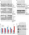
Protein expression analysis in NFKBIE-deleted versus WT CLL. (A) Protein expression profiles of IκBα, IκBβ and IκBε, p65, and phospho-p65 in NFKBIE-deleted (n = 4) versus WT (n = 4) CLL by Western blot analysis. (B) Protein expression profiles of IκBα, IκBβ and IκBε, p65, and phospho-p65 in additional NFKBIE-deleted (n = 3) versus WT (n = 3) CLL by Western blot analysis. (C) Mean normalized protein expression values for IκBα, IκBβ and IκBε, p65, and phospho-p65 in NFKBIE-deleted (n = 7) versus WT CLL (n = 7). * indicates P < 0.05, whereas *** indicates P < 0.001. For IκBβ, only four NFKBIE-deleted and four WT CLL cases were assessed. Error bars indicate standard error. (D) Western blot analysis of IκBε in CLL to identify the presence of a truncated IκBε protein. The membrane is overexposed. del, deleted.
Figure 4.
Figure 4.
Interactions between IκBs and p65 in CLL. (A) Co-IP to study the interaction between p65 and IκBε in NFKBIE-deleted (n = 3) versus WT (n = 2) CLL. The bottom panel indicates TCL from samples used in the co-IP assay. (B) Mean values for IκBε pull-down in NFKBIE-deleted versus WT CLL. (C) Proximity ligation assay to study the physical interaction between IκBα, IκBβ and IκBε, and p65 in six NFKBIE WT CLL patients. Interactions were assessed in unstimulated (U), αIgM-stimulated, and CD40L-stimulated cells. For IκBα and IκBε, six NFKBIE WT CLL patients were analyzed, whereas for IκBβ only four NFKBIE WT CLL patients were assessed. (D) The interaction between IκBε and the transcription factor p65 in six NFKBIE WT CLL patients and six NFKBIE-deleted patients. Interactions were again assessed in unstimulated (U), αIgM-stimulated, and CD40L-stimulated cells. Error bars indicate standard error. (E and F) Fluorescent microscope images of the interaction between IκBε and the transcription factor p65 in cells from a NFKBIE WT CLL patient (E) and an NFKBIE-deleted CLL patient (F). Blue color indicates cell nuclei, whereas each red dot represents a single interaction. Bars, 20 µm.
Figure 5.
Figure 5.
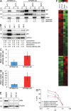
Functional analysis of the NFKBIE deletion in CLL. (A) Cytoplasmic (C) and nuclear (N) expression of p50, p65, and PARP in CLL patients. For the nuclear fraction, the expression ratio to PARP is provided. (B) Expression levels for p50, p65, and GAPDH in TCLs for the same patient. Normalized nuclear expression for p50 and p65 is provided. (C and D) Mean normalized nuclear expression of p50 (C) and p65 (D) in CLL. Error bars indicate standard error. (E) Western blot showing IκBε expression for the mock-transfected cells as well as the two independent knockdown clones (KD c1 and c2) in the HG3 cell line. All samples were run on the same gel but not in adjacent lanes and have therefore been quantified using the same exposure time. (F) Gene expression patterns in two independent CLL HG3 cell lines (c1 and c2) with partial knockdown (KD) of IκBε compared with the mock-transfected and untransfected (UT) HG3 cell lines. Genes that showed at least 50% difference in both c1 and c2 compared with either Mock or UT were selected. The list of genes is provided in Table S12. (G) Dose–response curves to the BTK inhibitor ibrutinib in primary CLL samples with WT NFKBIE (n = 4) and deleted NFKBIE (n = 4). Data were normalized against ibrutinib-naive CLL cells in culture. In NFKBIE WT cases, borderline significance was observed when comparing αIgM-stimulated versus unstimulated cells (P = 0.07 at 0.1 µM concentration, P = 0.06 at 1 µM concentration, and P = 0.05 at 2.5 µM concentration). Error bars indicate standard error.

Comment in

References

    1. Agathangelidis A., Darzentas N., Hadzidimitriou A., Brochet X., Murray F., Yan X.J., Davis Z., van Gastel-Mol E.J., Tresoldi C., Chu C.C., et al. . 2012. Stereotyped B-cell receptors in one-third of chronic lymphocytic leukemia: a molecular classification with implications for targeted therapies. Blood. 119:4467–4475. 10.1182/blood-2011-11-393694 - DOI - PMC - PubMed
    1. Alves B.N., Tsui R., Almaden J., Shokhirev M.N., Davis-Turak J., Fujimoto J., Birnbaum H., Ponomarenko J., and Hoffmann A.. 2014. IκBε is a key regulator of B cell expansion by providing negative feedback on cRel and RelA in a stimulus-specific manner. J. Immunol. 192:3121–3132. 10.4049/jimmunol.1302351 - DOI - PMC - PubMed
    1. Andrews N.C., and Faller D.V.. 1991. A rapid micropreparation technique for extraction of DNA-binding proteins from limiting numbers of mammalian cells. Nucleic Acids Res. 19:2499 10.1093/nar/19.9.2499 - DOI - PMC - PubMed
    1. Baliakas P., Hadzidimitriou A., Sutton L.-A., Minga E., Agathangelidis A., Nichelatti M., Tsanousa A., Scarfò L., Davis Z., Yan X.-J., et al. . 2014. Clinical effect of stereotyped B-cell receptor immunoglobulins in chronic lymphocytic leukaemia: a retrospective multicentre study. The Lancet Haematology. 1:e74–e84. 10.1016/S2352-3026(14)00005-2 - DOI - PubMed
    1. Baliakas P., Hadzidimitriou A., Sutton L.A., Rossi D., Minga E., Villamor N., Larrayoz M., Kminkova J., Agathangelidis A., Davis Z., et al. . European Research Initiative on CLL (ERIC). 2015. Recurrent mutations refine prognosis in chronic lymphocytic leukemia. Leukemia. 29:329–336. 10.1038/leu.2014.196 - DOI - PubMed

Publication types

MeSH terms