

Phagocytosis (cannibalism) of apoptotic neutrophils by tumor cells in gastric micropapillary carcinomas
- PMID: 25987778
- PMCID: PMC4427677
- DOI: 10.3748/wjg.v21.i18.5548
Phagocytosis (cannibalism) of apoptotic neutrophils by tumor cells in gastric micropapillary carcinomas
Abstract
Aim: To identify those with a micropapillary pattern, ascertain relative frequency and document clinicopathological characteristics by reviewing gastric carcinomas.
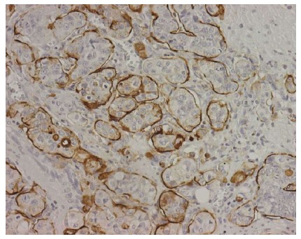
Methods: One hundred and fifty-one patients diagnosed with gastric cancer who underwent gastrectomy were retrospectively studied and the presence of a regional invasive micropapillary component was evaluated by light microscopy. All available hematoxylin-eosin (HE)-stained slides were histologically reviewed and 5 tumors were selected as putative micropapillary carcinoma when cancer cell clusters without a vascular core within empty lymphatic-like space comprised at least 5% of the tumor. Tumor tissues from these 5 invasive gastric carcinomas were immunostained using an anti-mucin 1 (MUC1) antibody (clone MA695) to detect the characteristic inside-out pattern and with D2-40 antibody to determine the presence of intratumoral lymph vessels. Detection of intraepithelial neutrophil apoptosis was evaluated in consecutive histological tissue sections by three independent methods, namely light microscopy with HE staining, the conventional terminal deoxynucleotidyl transferase-mediated dUTP-biotin nick end-labeling (TUNEL) method and immunohistochemistry for activated caspase-3 (clone C92-605).
Results: Among 151 gastric cancers resected for cure, 5 (3.3%) were adenocarcinomas with a micropapillary component. Four of the patients died of disease from 6 to 23 mo and one patient was alive with metastases at 9 mo. All patients had advanced-stage cancer (≥ pT2) and lymph node metastasis. Positive MUC1 immunostaining on the stroma-facing surface (inside-out pattern) of the carcinomatous cluster cells, together with negative immunostaining for D2-40 in the cells limiting lymphatic-like spaces, confirmed the true micropapillary pattern in these gastric neoplasms. In all five cases, several micropapillae were infiltrated by neutrophils. HE staining, TUNEL assay and immunostaining for caspase-3 demonstrated apoptotic neutrophils within cytoplasmic vacuoles of tumor cells. These data suggest phagocytosis (cannibalism) of apoptotic neutrophils by micropapillary tumor cells. Tumor cell cannibalism is usually found in aggressive tumors with anaplastic morphology. Our data extend these observations to gastric micropapillary carcinoma: a tumor histotype analogously characterized by aggressive behavior and poor prognosis. The results are of interest because they raise the intriguing possibility that neutrophil cannibalism by tumor cells may be one of the mechanisms favoring tumor growth in gastric micropapillary carcinomas.
Conclusion: This is the first study showing phagocytosis (cannibalism) of apoptotic neutrophils by tumor cells in gastric micropapillary carcinomas.
Keywords: Caspase-3; Gastric cancer; Micropapillary pattern; Mucin 1; TUNEL assay.
Figures


References
-
- Siriaunkgul S, Tavassoli FA. Invasive micropapillary carcinoma of the breast. Mod Pathol. 1993;6:660–662. - PubMed
-
- Amin MB, Ro JY, el-Sharkawy T, Lee KM, Troncoso P, Silva EG, Ordóñez NG, Ayala AG. Micropapillary variant of transitional cell carcinoma of the urinary bladder. Histologic pattern resembling ovarian papillary serous carcinoma. Am J Surg Pathol. 1994;18:1224–1232. - PubMed
-
- Amin MB, Tamboli P, Merchant SH, Ordóñez NG, Ro J, Ayala AG, Ro JY. Micropapillary component in lung adenocarcinoma: a distinctive histologic feature with possible prognostic significance. Am J Surg Pathol. 2002;26:358–364. - PubMed
-
- Michal M, Skálová A, Mukensnabl P. Micropapillary carcinoma of the parotid gland arising in mucinous cystadenoma. Virchows Arch. 2000;437:465–468. - PubMed
MeSH terms
Substances
LinkOut - more resources
Full Text Sources
Other Literature Sources
Medical
Research Materials
Miscellaneous
