

Chemokine RANTES/CCL5 as an unknown link between wound healing in the jawbone and systemic disease: is prediction and tailored treatments in the horizon?
- PMID: 25987906
- PMCID: PMC4435812
- DOI: 10.1186/s13167-015-0032-4
Chemokine RANTES/CCL5 as an unknown link between wound healing in the jawbone and systemic disease: is prediction and tailored treatments in the horizon?
Abstract
Background: This research elucidates the question of whether common and widespread dental procedures (DP) like root filling (RF) and the removal of wisdom teeth (WT) contribute to chronic inflammation in the jawbone. Dentists, in carrying out these DP, can set off defective wound healing in the jawbone in ignorance of its connection to inflammatory mediators and the possibility of it being a hidden cause of chronic systemic diseases (SYD).
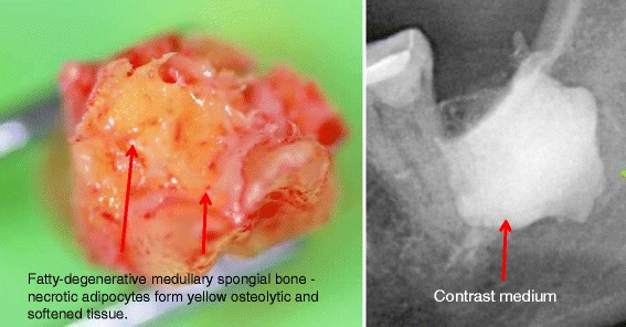
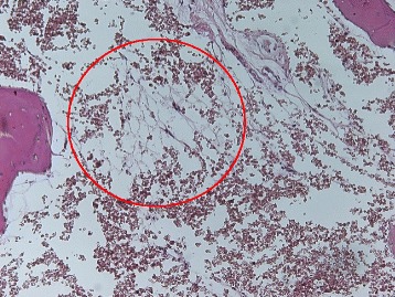
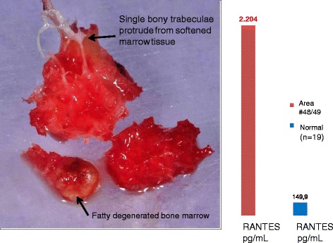
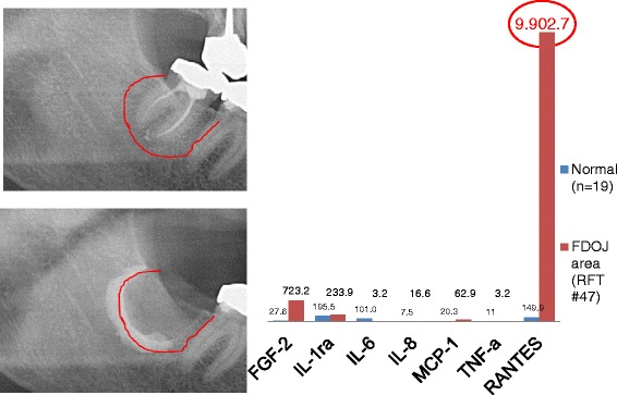
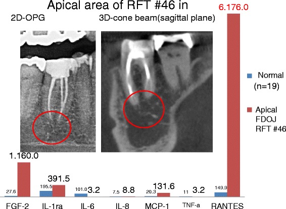
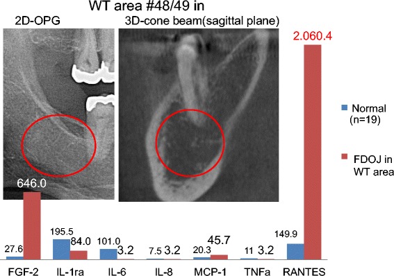
Materials and methods: We examined samples of the jawbone for seven cytokines by multiplex analysis in three groups of jawbone areas. In order to clarify systemic interrelations, specimens from 16 patients were analyzed in areas of former surgery in the retromolar wisdom tooth area; specimens from 16 patients were analyzed in the jawbone, apically of teeth with RF; and specimens from 19 patients were of the healthy jawbone. Each of the retromolar and the apical jawbone samples showed clinically fatty degenerated and osteonecrotic medullary changes.
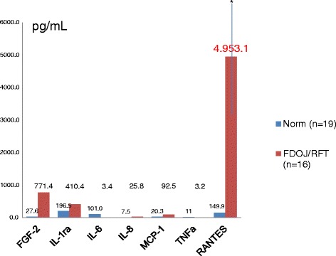
Results: All fatty necrotic and osteolytic jawbone (FDOJ) samples showed regulated on activation, normal T-cell expressed and secreted (RANTES) and fibroblast growth factor (FGF)-2 as the only extremely overexpressed cytokines. FDOJ cohorts showed a 30-fold mean overexpression of RANTES and a 20-fold overexpressed level of FGF-2 when compared to healthy controls.
Conclusions: As RANTES is discussed in the literature as a possible contributor to inflammatory diseases, and though it might have oncogenic effects, we hypothesize that FDOJ in areas of improper and incomplete wound healing in the jawbone might act as hyperactivated signaling pathways, while serving as an unknown source of "silent inflammation". Because of the wide range of RANTES in immune diseases, treating FDOJ can cover many potential prediction or prognosis of individual outcomes.
Keywords: Chronic inflammation in the jawbone; Fatty necrotic osteolytic jawbone; Hyperactivated signaling pathways; Predictive preventive personalized medicine; RANTES/CCL5.
Figures

References
-
- Townsend MJ, McKenzie AN. Unravelling the net? Cytokines and diseases. J Cell Sci. 2000;113(Pt 20):3549–50.
-
- Imbeau J. Introduction to through-transmission alveolar ultrasonography (TAU) in dental medicine. Cranio. 2005;23(2):100–12. - PubMed
-
- Ono K. Symposium: recent advances in avascular osteonecrosis. Clin Orthop. 1992;277:2.
LinkOut - more resources
Full Text Sources
Other Literature Sources
Research Materials
