. 2015 Mar 11:2:7.
doi: 10.3389/fmolb.2015.00007. eCollection 2015.

Pyroglutamyl-N-terminal prion protein fragments in sheep brain following the development of transmissible spongiform encephalopathies

Affiliations

Pyroglutamyl-N-terminal prion protein fragments in sheep brain following the development of transmissible spongiform encephalopathies

Adriana Gielbert et al. Front Mol Biosci. .

Abstract

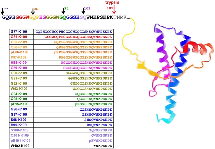
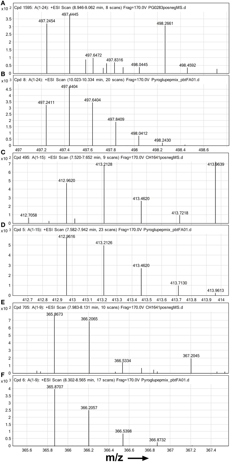
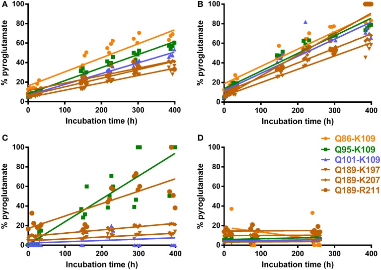
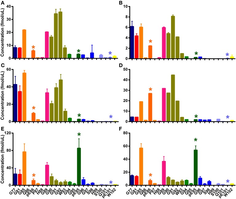
Protein misfolding, protein aggregation and disruption to cellular proteostasis are key processes in the propagation of disease and, in some progressive neurodegenerative diseases of the central nervous system, the misfolded protein can act as a self-replicating template or prion converting its normal isoform into a misfolded copy of itself. We have investigated the sheep transmissible spongiform encephalopathy, scrapie, and developed a multiple selected reaction monitoring (mSRM) mass spectrometry assay to quantify brain peptides representing the "ragged" N-terminus and the core of ovine prion protein (PrP(Sc)) by using Q-Tof mass spectrometry. This allowed us to identify pyroglutamylated N-terminal fragments of PrP(Sc) at residues 86, 95 and 101, and establish that these fragments were likely to be the result of in vivo processes. We found that the ratios of pyroglutamylated PrP(Sc) fragments were different in sheep of different breeds and geographical origin, and our expanded ovine PrP(Sc) assay was able to determine the ratio and allotypes of PrP accumulating in diseased brain of PrP heterozygous sheep; it also revealed significant differences between N-terminal amino acid profiles (N-TAAPs) in other types of ovine prion disease, CH1641 scrapie and ovine BSE. Variable rates of PrP misfolding, aggregation and degradation are the likely basis for phenotypic (or strain) differences in prion-affected animals and our mass spectrometry-based approach allows the simultaneous investigation of factors such as post-translational modification (pyroglutamyl formation), conformation (by N-TAAP analysis) and amino-acid polymorphisms (allotype ratio) which affect the kinetics of these proteostatic processes.

Keywords: amyloid disease; mass spectrometry; prions; pyroglutamate; strain differentiation.


Figures

Figure 1
Figure 1
Ovine prion protein model showing the amino acid sequence in the partially protease-sensitive region. Selected PK sites and the tryptic cleavage site are pointed out by the arrows. The resulting N-TAAP peptides and their N-terminal pyroglutamyl analogs where applicable are shown in the accompanying table, together with the sequence references used. The ovine PrP structure shown is for illustration only and was captured from Protein Data Bank entry 1Y2S (ovine PrP fragment V124-S234) using GLmol – Molecular Viewer (Version 0.47).
Figure 2
Figure 2
High resolution mass spectra of ovine PrP peptides with pyroglutamate N-termini. Mass spectra are shown zoomed in on the isotope distribution of (A,B) quintuply protonated pEPHGGGWGQGGSHSQWNKPSKPK (pE86-K109) (A) suspected in PrPSc digest from classical scrapie infected brain sample and (B) synthetic standard; (C,D) quadruply protonated pEGGSHSQWNKPSKPK (pE95-K109) and (E,F) triply protonated pEWNKPSKPK (C,E) suspected in PrPSc digest from an experimental CH1641 scrapie brain sample and (D,F) synthetic standards. Chip-HPLC Q-Tof mass spectra were obtained by integrating the total MS signal over the full width at base of the chromatographic peak of the peptide. Spectra (B,D,F) were based on 100 fmol on column of each standard.
Figure 3
Figure 3
Conversion rates of N-terminal glutamine to pyroglutamate for six PrP peptides. Chip-HPLC Q-Tof mass spectrometry was used. Repeat analyses of N-terminal glutamine peptide mixtures in water, PTB-FA and PBT-HCl, spaced by buffer blanks, were run and the injection times recorded as described in Materials and Methods. (A) Post-trypsin buffer (PTB) to which 0.5 volume 5% formic acid was added as by PrPSc digest preparation protocol (final pH 3.0) (B) PTB plus 0.5 volume 5% HCl (pH 1.0) (C) PTB + 0.5 volume water (pH 8.0) (D) peptides dissolved in pure water (pH 7.0). Q-pE conversion rates calculated from these plots are given in Table 1.
Figure 4
Figure 4
N-TAAPs from ovine TSEs. Graphs show calculated concentrations (±SD) of N-TAAP peptides determined by chip-HPLC SRM mass spectrometry of digest preparations from the brain stem of individual animals. (A,B) Classical scrapie: VRQ/VRQ Swaledale, neutered male, homebred; (C,D) Experimental CH1641 scrapie: AHQ/AHQ Cheviot, neutered male; (E,F) Experimental BSE: ARQ/ARQ Romney. (A,C,E) without proteinase K treatment; (B,D,F) with proteinase K treatment. Asterisks are used to point out the peptides with pyroglutamyl N-termini.
Figure 5
Figure 5
N-TAAPs from natural classical scrapie samples. Graphs show calculated concentrations (±SD) of N-TAAP peptides determined by chip-HPLC SRM mass spectrometry of digest preparations from the brain stem of individual animals, obtained by processing 2 × 1.75 ml of 10% homogenate with PK treatment. (A) ARQ/ARQ Swaledale, neutered male, homebred; (B) VRQ/VRQ Swaledale, female, homebred; (C) ARH/VRQ Texel, female, purchased into flock from farm A; (D) ARQ/VRQ white-faced Dartmoor, female, homebred (E,F) ARQ/ARQ Vendeen, female, purchased into flock from farm B. Asterisks are used to point out the peptides with pyroglutamyl N-termini.
Figure 6
Figure 6
N-TAAPs from experimental ovine TSEs. Graphs show calculated concentrations (±SD) of N-TAAP peptides determined by chip-HPLC SRM mass spectrometry of digest preparations from the brain stem of individual animals, obtained by processing 2 × 1.75 ml of 10% homogenate. All animals were neutered males of the Cheviot breed, AHQ/AHQ genotype. (A,B) PK treated CH1641 scrapie (Coombelands farm, Surrey); (C,D) PK treated ovine BSE (ADAS Drayton, Warwicksire); (E,F) ovine BSE samples corresponding to (C,D) without PK treatment. Asterisks are used to point out the peptides with pyroglutamyl N-termini.

References

    1. Baldwin M. A. (2001). Mass spectrometric analysis of prion proteins. Adv. Protein Chem. 57, 29–54 10.1016/S0065-3233(01)57017-5 - DOI - PubMed
    1. Baldwin M. A., Falick A. M., Gibson B. W., Prusiner S. B., Stahl N., Burlingame A. L. (1990). Tandem mass spectrometry of peptides with N-terminal glutamine. Studies on a prion protein peptide. J. Am. Soc. Mass Spectrom. 1, 258–264 10.1016/1044-0305(90)85043-L - DOI
    1. Bruce M. E. (1993). Scrapie strain variation and mutation. Br. Med. Bull. 49, 822–838. - PubMed
    1. Carr S. A., Biemann K. (1984). Identification of posttranslationally modified amino acids in proteins by mass spectrometry, in Methods in Enzymology, eds Wold F., Moldave K. (Orlando, FL: Elsevier; ), 29–58. - PubMed
    1. Chelius D., Jing K., Lueras A., Rehder D. S., Dillon T. M., Vizel A., et al. . (2006). Formation of pyroglutamic acid from N-terminal glutamic acid in immunoglobulin gamma antibodies. Anal. Chem. 78, 2370–2376. 10.1021/ac051827k - DOI - PubMed