. 2015 Aug:85:37-47.
doi: 10.1016/j.yjmcc.2015.05.007. Epub 2015 May 16.

Losartan treatment attenuates tumor-induced myocardial dysfunction

Affiliations

Losartan treatment attenuates tumor-induced myocardial dysfunction

Sarah C W Stevens et al. J Mol Cell Cardiol. 2015 Aug.

Abstract

Fatigue and muscle wasting are common symptoms experienced by cancer patients. Data from animal models demonstrate that angiotensin is involved in tumor-induced muscle wasting, and that tumor growth can independently affect myocardial function, which could contribute to fatigue in cancer patients. In clinical studies, inhibitors of angiotensin converting enzyme (ACE) can prevent the development of chemotherapy-induced cardiovascular dysfunction, suggesting a mechanistic role for the renin-angiotensin-aldosterone system (RAAS). In the present study, we investigated whether an angiotensin (AT) 1-receptor antagonist could prevent the development of tumor-associated myocardial dysfunction.

Methods and results: Colon26 adenocarcinoma (c26) cells were implanted into female CD2F1 mice at 8weeks of age. Simultaneously, mice were administered Losartan (10mg/kg) daily via their drinking water. In vivo echocardiography, blood pressure, in vitro cardiomyocyte function, cell proliferation assays, and measures of systemic inflammation and myocardial protein degradation were performed 19days following tumor cell injection. Losartan treatment prevented tumor-induced loss of muscle mass and in vitro c26 cell proliferation, decreased tumor weight, and attenuated myocardial expression of interleukin-6. Furthermore, Losartan treatment mitigated tumor-associated alterations in calcium signaling in cardiomyocytes, which was associated with improved myocyte contraction velocity, systolic function, and blood pressures in the hearts of tumor-bearing mice.

Conclusions: These data suggest that Losartan may mitigate tumor-induced myocardial dysfunction and inflammation.

Keywords: Calcium signaling; Cancer cachexia; Cardiomyocyte; Cardiovascular function; Losartan.


Conflict of interest statement

Conflict of Interest

None declared

Figures

Figure 1
Figure 1
Gene expression of MAFbx (A), Bnip3 (B) and IL-6 (C) in cardiac tissue of tumor-bearing and Losartan-treated mice. Gene expression levels were determined using RT-PCR and are normalized to GAPDH expression. A, MAFbx, an indicator of ubiquitin proteasome function; B, Bnip3, an indicator of cellular autophagy; C, IL-6, an inflammatory cytokine. Data were analyzed using two-way ANOVA and Bonferroni post hoc pairwise comparisons. * p<0.05 compared to respective control. n=6 control/sham, n=5 tumor/sham, n=6 control/LOS, n=7 tumor/LOS.
Figure 2
Figure 2
Plasma pro-inflammatory cytokine levels in tumor-bearing and Losartan-treated mice. Cytokine levels were determined using the Pro-inflammatory 7-Plex Ultra-Sensitive Kit. Data were analyzed using two-way ANOVA (tumor growth and Losartan treatment) and Bonferroni post hoc pairwise comparisons. * p<0.05 compared to respective control, n=10.
Figure 3
Figure 3
Representative M-mode in vivo echocardiographic images obtained at the mid-papillary muscle level of tumor-bearing and Losartan-treated mice. Images include electrocardiogram and respiratory traces at the bottom.
Figure 4
Figure 4
In vivo myocyte function in tumor-bearing and Losartan-treated mice. A, percent peak sarcomere shortening (Sarcomere BL). B, time to 90 percent of peak shortening (TPS90). C, time to 90 percent relengthening (TR90). D, velocity of myocyte shortening. E, velocity of myocyte relengthening. Data were analyzed using two-way ANOVA (tumor growth and Losartan treatment) and Bonferroni post hoc pairwise comparisons. * p<0.05 compared to respective control. Myocyte cell numbers: n=70 cells/12 mice control/sham, n=97 cells/17 mice tumor/sham, n=45 cells/4 mice control/LOS, n=61 cells/8 mice tumor/LOS.
Figure 5
Figure 5
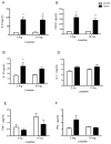
Cardiomyocyte calcium signaling in tumor-bearing and Losartan-treated mice. A, amplitude of stimulated Ca2+ transients. B, amplitude of calcium sparks. C, amplitude of caffeine induced Ca2+ release. D, Integration under the caffeine-induced Ca2+ release curve as an index of SR Ca2+ concentration. Data analyzed using two-way ANOVA (tumor growth, Losartan treatment), followed by Bonferroni post hoc pairwise comparisons. * p<0.05 compared to respective control. Myocyte cell numbers for calcium analysis: n=27 cells/11 mice control/sham, n=40 cells/14 mice tumor/sham, n=29 cells/4 mice control/LOS, n=52 cells/8 mice tumor/LOS.
Figure 6
Figure 6
Oxidative stress in ventricular tissue of tumor-bearing and Losartan-treated mice. The ratio of GSH/GSSG was used to approximate oxidative stress in left ventricular tissue. Data analyzed using two-way ANOVA (tumor growth, Losartan treatment), followed by Bonferroni post hoc pairwise comparisons. No significance was observed with Losartan treatment. n=5.
Figure 7
Figure 7
Angiotensin II Protein levels in mouse serum of tumor-bearing and Losartan treated mice 18 days after tumor cell or sham injection. Data were analyzed using two-way ANOVA (tumor growth, Losartan treatment), followed by Bonferroni post hoc pairwise comparisons. * p<0.01 compared to control, n=4.
Figure 8
Figure 8
Effects of Losartan on c26 Adenocarcinoma cell proliferation. Cells were cultured with 1 mM/L, 10 mM/L or without Losartan for 3 days. Data were analyzed using one-way ANOVA. Losartan significantly inhibited the proliferation of c26 Adenocarcinoma cell at both concentrations. p<0.001, n=6.

References

    1. Douglas E, McMillan DC. Towards a simple objective framework for the investigation and treatment of cancer cachexia: The Glasgow Prognostic Score. Cancer treatment reviews. 2014;40:685–91. - PubMed
    1. Tian M, Asp ML, Nishijima Y, Belury MA. Evidence for cardiac atrophic remodeling in cancer-induced cachexia in mice. International journal of oncology. 2011;39:1321–6. - PubMed
    1. Xu H, Crawford D, Hutchinson KR, Youtz DJ, Lucchesi PA, Velten M, et al. Myocardial dysfunction in an animal model of cancer cachexia. Life sciences. 2011;88:406–10. - PMC - PubMed
    1. Zuppinger C, Timolati F, Suter TM. Pathophysiology and diagnosis of cancer drug induced cardiomyopathy. Cardiovascular toxicology. 2007;7:61–6. - PubMed
    1. Tian M, Nishijima Y, Asp ML, Stout MB, Reiser PJ, Belury MA. Cardiac alterations in cancer-induced cachexia in mice. International journal of oncology. 2010;37:347–53. - PubMed

Publication types

MeSH terms