. 2015 Sep;146(1):89-99.
doi: 10.1111/imm.12484. Epub 2015 Jun 25.

Differential dependence on nuclear factor-κB-inducing kinase among natural killer T-cell subsets in their development

Affiliations

Differential dependence on nuclear factor-κB-inducing kinase among natural killer T-cell subsets in their development

Haruka Noma et al. Immunology. 2015 Sep.

Abstract

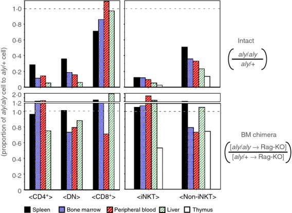
Natural killer T cells (NKT cells) are comprised of several subsets. However, the possible differences in their developmental mechanisms have not been fully investigated. To evaluate the dependence of some NKT subpopulations on nuclear factor-κB-inducing kinase (NIK) for their generation, we analysed the differentiation of NKT cells, dividing them into subsets in various tissues of alymphoplasia (aly/aly), a mutant mouse strain that lacks functional NIK. The results indicated that the efficient differentiation of both invariant NKT (iNKT) and non-iNKT cells relied on NIK expression in non-haematopoietic cells; however, the dependence of non-iNKT cells was lower than that of iNKT cells. Especially, the differentiation of CD8(+) non-iNKT cells was markedly resistant to the aly mutation. The proportion of two other NKT cell subsets, NK1.1(+) γδ T cells and NK1.1(-) iNKT cells, was also significantly reduced in aly/aly mice, and this defect in their development was reversed in wild-type host mice given aly/aly bone marrow cells. In exerting effector functions, NIK in NKT-αβ cells appeared dispensable, as NIK-deficient NKT-αβ cells could secrete interleukin-4 or interferon-γ and exhibit cytolytic activity at a level comparable to that of aly/+ NKT-αβ cells. Collectively, these results imply that the NIK in thymic stroma may be critically involved in the differentiation of most NKT cell subsets (although the level of NIK dependence may vary among the subsets), and also that NIK in NKT-αβ cells may be dispensable for their effector function.

Keywords: differentiation; natural killer T cells; nuclear factor-κB-inducing kinase.


Figures

Figure 1
Figure 1
The proportion of NK1.1+ T-cell receptor-αβ (TCR-αβ) T cells in various tissues in aly/aly or aly/+ mice. The leucocytes were collected from the indicated tissues of aly/aly or aly/+ mice (= 6) and the proportions of NK1.1+ αβ T cells in them were examined by flow cytometry. The percentages of NK1.1+ αβ T cells in total TCR-αβ+ cells are depicted, and the bar in each graph indicates the average.
Figure 2
Figure 2
Analyses of NK1.1+ αβ T cells in bone marrow from aly/aly or aly/+ mice. Bone marrow cells from aly/aly or aly/+ mice were analysed for CD4 or CD8 expression and for binding of α-GalCer/CD1d dimer. (a) The representative FACS profiles are shown. In the left panels, the expression of T-cell receptor-αβ (TCR-αβ) and NK1.1 on total leucocytes is shown where the numbers indicate the percentage of NK1.1+ αβ T cells. The NK1.1+ αβ T cells were gated to analyse their co-receptor expression (the middle panels). In the right panels, total TCR-αβ T cells were analysed for NK1.1 expression and α-GalCer/CD1d binding. (b, c) The percentages of NK1.1+ αβ T cells of the indicated phenotypes from bone marrow of aly/aly or aly/+ mice are shown.
Figure 3
Figure 3
The differential requirement for nuclear factor-κB-inducing kinase (NIK) among the subsets of NK1.1+ αβ T cells in their optimal generation. The NK1.1+ αβ T cells from thymus, spleen, peripheral blood and liver of aly/aly or aly/+ mice were analysed by flow cytometry, and the results are shown in the same manner as in Fig.2.
Figure 4
Figure 4
Recovery of nuclear factor-κB-inducing kinase (NIK) -deficient NK1.1+, αβ T-cell generation in bone marrow chimera of wild-type hosts injected with aly/aly donor cells (upper panels). The average number of aly/aly natural killer T (NKT) cells obtained from the indicated organs were divided by the aly/+ NKT cell number, and the proportion of aly/aly against aly/+ are shown (lower panels). The bone marrow chimeric mice were prepared by transfusing aly/aly or aly/+ bone marrow cells into irradiated RAG2-KO hosts, to evaluate the impact of NIK-impairment in non-haematopoietic cells on NK1.1+ αβ T-cell development. The proportion of aly/aly NK1.1+ αβ T cells against aly/+ cells in chimeric mice are shown. The average numbers from four to six mice for each group were used to calculate the proportion.
Figure 5
Figure 5
The proper generation of NK1.1+ γδ T cells or NK1.1 invariant natural killer T (iNKT) cells also requires nuclear factor-κB-inducing kinase (NIK) expression in non-haematopoietic cells. The thymocytes and splenocytes from aly/aly or aly/+ mice, or [aly/aly → RAG2-KO] or [aly/+ → RAG2-KO] chimera mice were analysed for the expression of NK1.1 and T-cell receptor-γδ (TCR-γδ), and for α-GalCer/CD1d-binding. (a) Representative FACS profiles of NK1.1 and TCR-γδ expression on the splenic T cells from aly/aly or aly/+ mice are demonstrated. The numbers in the panels indicate the percentage of NK1.1+ γδ T cells in CD3+ cells (upper panels). The percentage of NK1.1+ γδ T cells in CD3hi+ thymocytes (left) or of those in CD3+ splenocytes (right) from aly/aly or aly/+ mice, or from bone marrow (BM) chimera are shown (lower panels). Six mice were analysed for each group. (b) A representative set of FACS analyses of the splenic T cells from aly/aly or aly/+ mice for the expression of NK1.1 and binding of α-GalCer/CD1d are demonstrated in upper panels. In the lower panels, the average percentage of NK1.1 iNKT cells from six mice of aly/aly or aly/+ mice, or BM chimera are presented.
Figure 6
Figure 6
Normal expression of interleukin-7 (IL-7) or IL-15 mRNA in cortical thymic epithelial cells (TECs) from aly/aly mouse. The thymi from aly/aly or aly/+ mice were treated with Liberase and the cells were stained with antibodies to CD45, CD326 (EpCAM-1), or Ly51, and with Ulex Europaeus Agglutinin 1 (UEA-1), to sort cortical (cTECs) or medullary (mTECs). (a) The expression of CD45 and CD326 on total cells from thymus are shown in upper panels. The CD45 CD326+ TECs were further separated into Ly51+ cTECs and UEA-1-binding mTECs (lower panels). (b) Total RNA was extracted from FACS-sorted cTECs of aly/aly or aly/+ mice to quantify the mRNA of IL-7 or IL-15. (c) The mRNA of IL-7, IL-15, or IL-15Rα expressed in cTECs or mTECs from wild-type mice were compared. Relative amounts of each mRNA normalized with Hprt mRNA are displayed. Similar results were obtained from three or four independent experiments and a representative set of results is shown.
Figure 7
Figure 7
The nuclear factor-κB-inducing kinase (NIK) in NK1.1+ αβ T cells is dispensable for their function of cytokine production or cytolysis. The NK1.1+ αβ T cells were sorted from [aly/aly → RAG2-KO] or [aly/+ → RAG2-KO] chimera mice and their functions were investigated. (a) Either 100 or 50 000 of CD4+ or double-negative (DN) NK1.1+ αβ T cells, respectively, were stimulated in vitro with anti-CD3 antibody, coated on the surface of the wells at 10 μg/ml. After 20 hr, the concentration of interferon-γ (IFN-γ) or interleukin-4 (IL-4) in the supernatants was quantified by ELISA. The proportion of invariant natural killer T (iNKT) cells in the CD4+ subset was 85·1 ± 2·7% for aly/+ donor mice, and 83·4 ± 3·3% for aly/aly donor mice. The iNKT proportion in DN cells was 61·7 ± 11·6% for aly/+ donor mice, and 60·2 ± 4·0% for aly/aly donor mice. (b) The NK1.1+ αβ T cells of indicated phenotype were sorted from bone marrow (BM) of aly/aly or aly/+ mice and used as effector cells in re-directed cell-mediated lysis assay to examine their cytolytic activity. The proportion of iNKT cells in CD8+, DN or CD4+ subsets in bone marrow were as follows: in CD8+ cells, 1·0 ± 0·6% for aly/+ donor mice, and 1·8 ± 1·5% for aly/aly donor mice; in DN cells, 8·5 ± 0·9% for aly/+ donor mice and 8·6 ± 2·9% for aly/aly donor mice; and in CD4+ cells, 62·3 ± 5·8% for aly/+ donor mice and 53·5 ± 11·0% for aly/aly donor mice.

Similar articles

Cited by

References

    1. Bendelac A, Savage PB, Teyton L. The biology of NKT cells. Annu Rev Immunol. 2007;25:297–336. - PubMed
    1. Kumar V, Delovitch TL. Different subsets of natural killer T cells may vary in their roles in health and disease. Immunology. 2014;142:321–36. - PMC - PubMed
    1. Watarai H, Sekine-Kondo E, Shigeura T, et al. Development and function of invariant natural killer T cells producing Th2- and Th17-cytokines. PLoS Biol. 2012;10:e1001255. - PMC - PubMed
    1. Lee YJ, Holzapfel KL, Zhu J, Jameson SC, Hogquist KA. Steady-state production of IL-4 modulates immunity in mouse strains and is determined by lineage diversity of iNKT cells. Nat Immunol. 2013;14:1146–54. - PMC - PubMed
    1. Michel ML, Mendes-da-Cruz D, Keller AC, Lochner M, Schneider E, Dy M, Eberl G, Leite-de-Moraes MC. Critical role of ROR-γt in a new thymic pathway leading to IL-17-producing invariant NKT cell differentiation. Proc Natl Acad Sci USA. 2008;105:19845–50. - PMC - PubMed

Publication types

MeSH terms