Review
. 2015 Aug:35:98-105.
doi: 10.1016/j.ceb.2015.04.015. Epub 2015 May 16.

The essential roles of transition fibers in the context of cilia

Affiliations
Review

The essential roles of transition fibers in the context of cilia

Qing Wei et al. Curr Opin Cell Biol. 2015 Aug.

Abstract

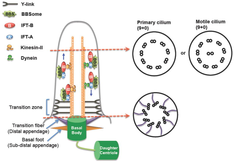
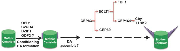
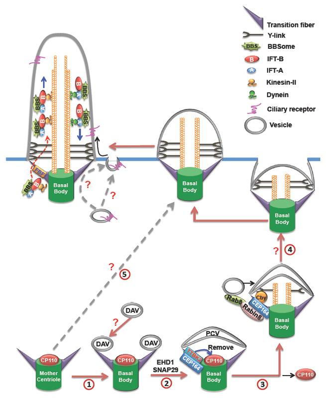
Once thought of as a vestigial organelle, the primary cilium is now recognized as a signaling hub for key cellular pathways in vertebrate development. The recent renaissance in cilia studies significantly improved our understanding of how cilia form and function, but little is known about how ciliogenesis is initiated and how ciliary proteins enter cilia. These important ciliary events require transition fibers (TFs) that are positioned at the ciliary base as symmetric nine-bladed propeller fibrous structures. Up until recently, TFs have been the most underappreciated ciliary structures due to limited knowledge about their molecular composition and function. Here, we highlight recent advances in our understanding of TF composition and the indispensable roles of TFs in regulating the initiation of ciliogenesis and the selective import of ciliary proteins.


Figures

Figure 1
Figure 1. Schematic diagram of a cilium
All cilia have a microtubule-based core structure, called the axoneme, which projects from the basal body and is tightly surrounded by the ciliary membrane. Based on the motility, cilia can be divided into primary cilia (non-motile cilia) and motile cilia. The axoneme of primary cilia typically consists of nine microtubule doublets (9+0), typical motile cilia have an extra pair of microtubule singlet in the center of the ring of nine outer doublets (9+2). The basal body is transformed from the mother centriole during ciliogenesis. At the ciliary base, there are two structurally distinct sub-regions: TFs and the TZ. TFs are analogous to DAs of the mother centriole and form a 9-bladed propeller-like structure linking the basal body to the ciliary membrane. Basal feet (analogous to sub-DAs) locate below TFs on the basal body. Above TFs is the TZ that is characterized by the Y-links connecting axoneme microtubules to the ciliary membrane. Extension, maintenance and function of cilia require intraciliary transport machinery IFT, which is composed of IFT-A, IFT-B, BBSome and motors. IFT moves bidirectionally along the ciliary axoneme to transport cargos into or out of cilia. TFs, transition fibers; TZ, transition zone; DAs, distal appendages; IFT, Intraflagellar transport; Sub-DAs, sub-distal appendages.
Figure 2
Figure 2. The assembly of distal appendages/transition fibers
OFD1, C2CD3, DZIP1, and ODF2 condition the distal end of the mother centriole for DA formation. Then different structural/functional components of DAs/TFs are recruited in a sequential manner. DAs, distal appendages; TFs, transition fibers.
Figure 3
Figure 3. The roles of transition fibers in the context of cilia
In some mammalian cell types, during the very early stage of ciliogenesis, small DAVs first dock to the DAs of the mother centriole through an unknown mechanism (1). EHD1 and SNAP29 then regulate the fusion of DAVs into the large PCV (2). During the formation of PCV, TTBK2 is recruited to TFs by CEP164 to remove microtubule cap protein CP110 to initiate axoneme elongation. Then the interaction between Rabin8/Rab8 complex and CEP164/Cby complex mediates the recruitment of more Rab8 positive vesicles to support membrane extension of the PCV (3). Meantime, the TZ starts to form, the basal body-PCV migrates to the plasma membrane, and then the PCV fuses with the cell membrane through an unknown mechanism (4). Lastly, IFT regulates the extension of the axoneme. FBF1 acts as the functional component on TFs to facilitate the ciliary import of assembled IFT complex. Polarized vesicle trafficking and exocytosis have been implicated in mediating ciliary cargos targeting to periciliary membrane, and then these cargos enter into cilia through lateral diffusion. But, whether TFs play a role in this process is not clear. In other cell types, an alternative ciliogenesis pathway (5) may be employed: the basal body directly docks to the plasma membrane independent of DAV/PCV route. TFs, transition fibers; TZ, transition zone; DAs, distal appendages; DAVs, distal appendage vesicles; PCV, primary ciliary vesicle.

References

    1. Gerdes JM, Davis EE, Katsanis N. The vertebrate primary cilium in development, homeostasis, and disease. Cell. 2009;137:32–45. - PMC - PubMed
    1. Goetz SC, Anderson KV. The primary cilium: a signalling centre during vertebrate development. Nat Rev Genet. 2010;11:331–344. - PMC - PubMed
    1. Hildebrandt F, Benzing T, Katsanis N. Ciliopathies. N Engl J Med. 2011;364:1533–1543. - PMC - PubMed
    1. Badano JL, Mitsuma N, Beales PL, Katsanis N. The ciliopathies: an emerging class of human genetic disorders. Annu Rev Genomics Hum Genet. 2006;7:125–148. - PubMed
    1. Baker K, Beales PL. Making sense of cilia in disease: the human ciliopathies. American journal of medical genetics Part C, Seminars in medical genetics. 2009;151C:281–295. - PubMed

Publication types