. 2015 Aug;172(16):4089-106.
doi: 10.1111/bph.13189. Epub 2015 Jun 26.

Pelitinib (EKB-569) targets the up-regulation of ABCB1 and ABCG2 induced by hyperthermia to eradicate lung cancer

Affiliations

Pelitinib (EKB-569) targets the up-regulation of ABCB1 and ABCG2 induced by hyperthermia to eradicate lung cancer

Kenneth K W To et al. Br J Pharmacol. 2015 Aug.

Abstract

Background and purpose: Pelitinib is a potent irreversible EGFR TK inhibitor currently in clinical trials for the treatment of lung cancer. Hyperthermia has been applied concomitantly with chemotherapy and radiotherapy to enhance treatment outcome. In this study, we investigated the ability of the combination of pelitinib with other conventional anticancer drugs to specifically target cancer cells with up-regulated efflux transporters ABCB1/ABCG2 after hyperthermia as a novel way to eradicate the cancer stem-like cells responsible for cancer recurrence.

Experimental approach: Alterations in intracellular topotecan accumulation, the efflux of fluorescent probe substrates, expression and ATPase activity of ABCB1/ABCG2 and tumoursphere formation capacity of side population (SP) cells sorted after hyperthermia were examined to elucidate the mechanism of pelitinib-induced chemosensitization.

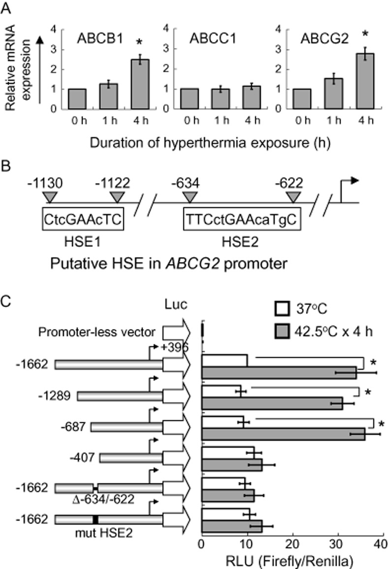
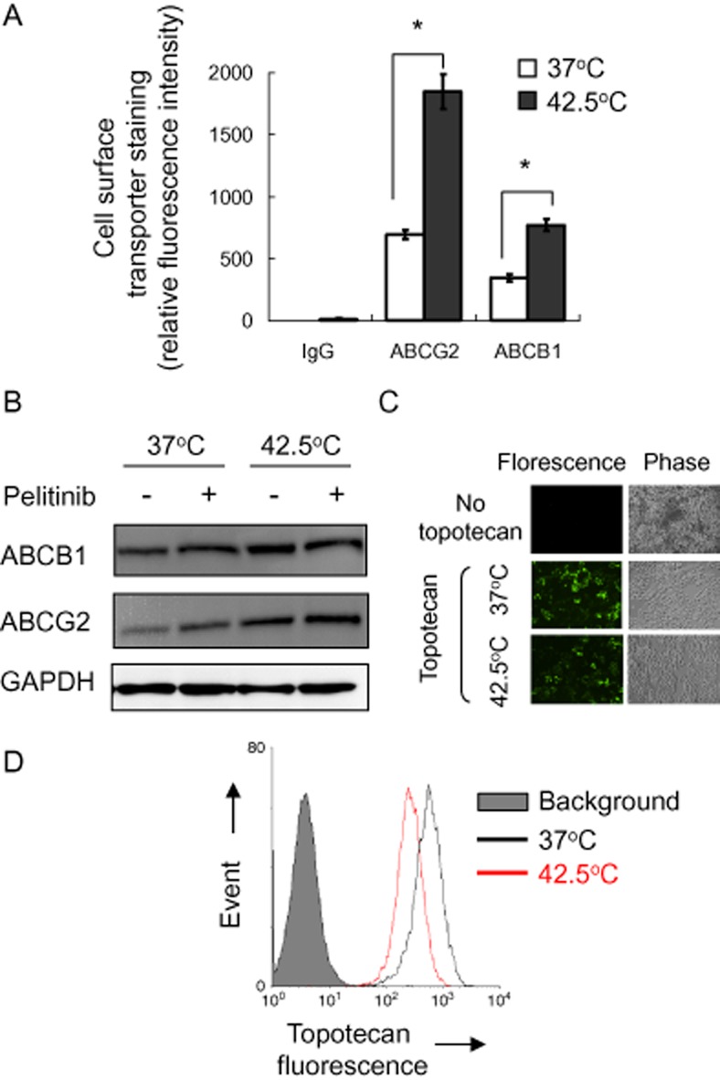
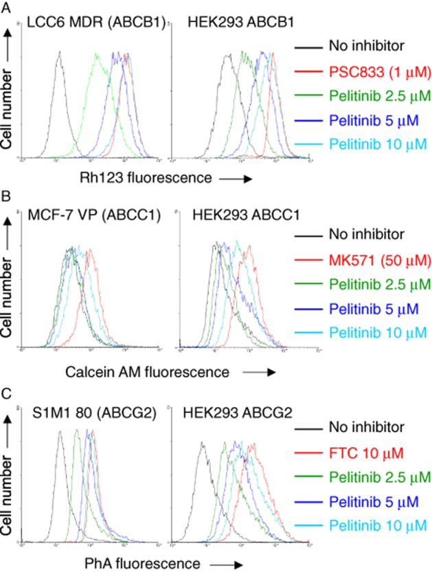
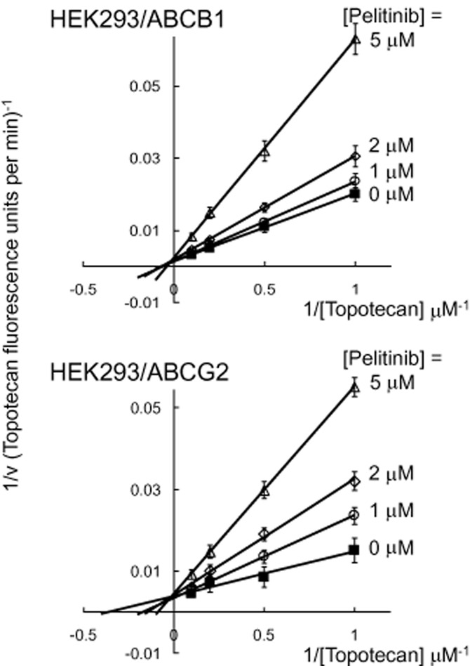
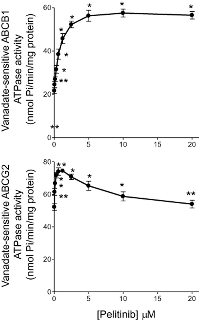
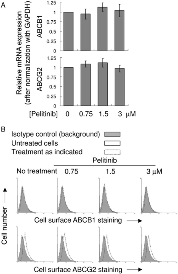
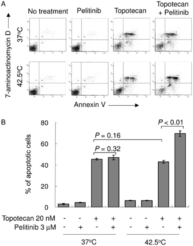
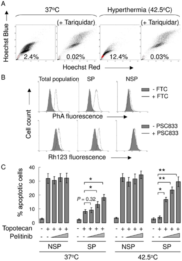
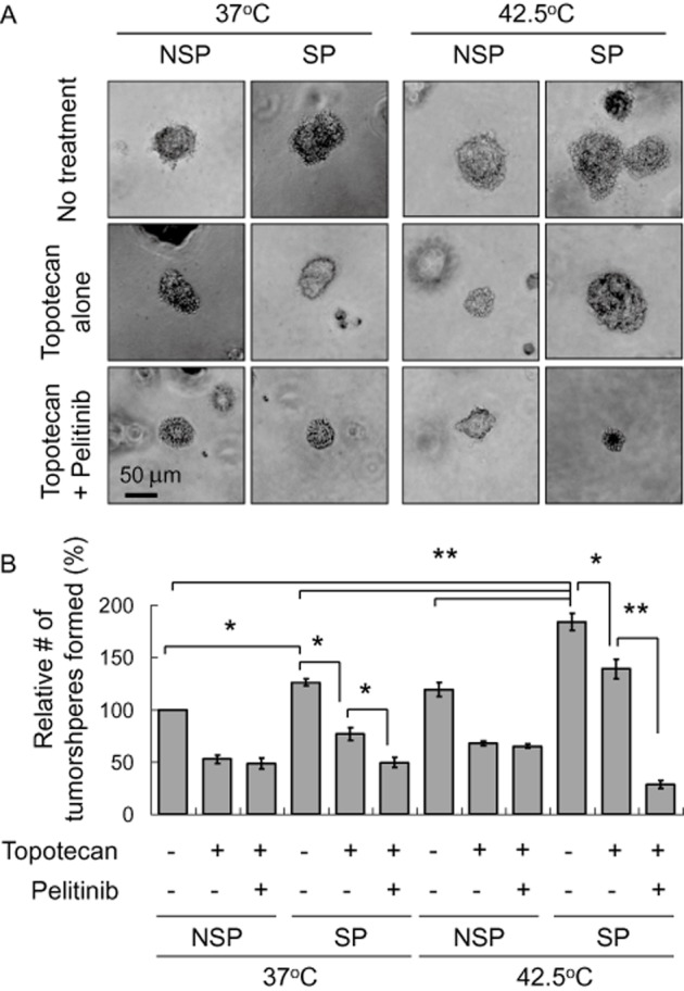
Key results: While pelitinib did not modulate ABCB1/ABCG2 expressions, the combination of pelitinib with transporter substrate anticancer drugs induced more marked apoptosis, specifically in cells exposed to hyperthermia. The flow cytometric assay showed that both ABCB1- and ABCG2-mediated drug effluxes were significantly inhibited by pelitinib in a concentration-dependent manner. The inhibition kinetics suggested that pelitinib is a competitive inhibitor of ABCB1/ABCG2, which is consistent with its ability to stimulate their ATPase activity. SP cells sorted after hyperthermia were found to be more resistant to anticancer drugs, presumably due to the up-regulation of ABCB1 and ABCG2. Importantly, pelitinib specifically enhanced the chemosensitivity but reduced the tumoursphere formation capacity of these SP cells.

Conclusions and implications: This study demonstrated a novel approach, exploiting drug resistance, to selectively kill cancer stem-like cells after hyperthermia.


Figures

Figure 1
Figure 1
Up-regulation of ABCB1 and ABCG2 by hyperthermia in A549 cells. (A) Real-time PCR analysis of ABCB1, ABCC1 and ABCG2 mRNA expression in A549 with or without hyperthermia treatment at 42.5°C for 1 or 4 h. Relative ABCB1, ABCC1 or ABCG2 transcript expression is shown after normalization with GAPDH. *P < 0.05, compared with cell culture at 37°C. (B) Schematic illustration of two putative HSEs (HSE1 and HSE2) in the ABCG2 promoter. The upper case letters represent the nucleotides matching the reported human consensus HSE sequence (TTCnnGAAnnTTC; n = any nucleotide; Akerfelt et al., 2010). (C) ABCG2 promoter luciferase reporter assay. Left, schematic representation of the 5′-deletion ABCG2 promoter constructs. The 5′-end of each of the constructs relative to the transcription start site (arrows) is indicated. The pGL3-basic (promoterless) vector, encoding firefly luciferase, was used to determine the basal levels. The construct (Δ-634/-622) was prepared with the putative HSE2 deleted from the full-length ABCG2 promoter, whereas the other construct (mut HSE2) was prepared with the putative HSE2 mutated from TTCctGAAcaTgC to AGTctAGCctGgA. Right, ABCG2 transcriptional activity in A549 cells transiently transfected with the various ABCG2 promoter constructs with or without hyperthermia treatment at 42.5°C for 4 h. The mean reporter activity ± SD [Firefly/Renilla luciferase units (RLU)] from three independent experiments is presented. *P < 0.05, compared with cell culture at 37°C.
Figure 2
Figure 2
Cell surface expression and functional analysis of ABCG2 and ABCB1 in A549 cells with or without hyperthermia (42.5°C) treatment. (A) Cell surface ABCG2 or ABCB1 staining of A549 cells with or without hyperthermia (42.5°C) pretreatment was quantified by subtracting the fluorescence signal from 5D3 or UIC2 labelling by that from the control IgG isotype antibody labelling. Mean ± SD from three independent experiments is shown. *P < 0.05, compared with cell culture at 37°C. (B) Western blot analysis of total ABCB1 or ABCG2 protein expression in A549 cells with or without 4 h of hyperthermia (42.5°C) treatment. It is also noted that pelitinib treatment did not alter the up-regulated ABCB1 or ABCG2 protein expression after hyperthermia (42.5°C) treatment. (C) Fluorescence microscopy images showing the steady state accumulation of a fluorescent ABCG2/ABCB1 substrate anticancer drug topotecan (drug incubation at 37°C × 60 min) in A549 cells with or without 4 h of hyperthermia pretreatment. Left panel, fluorescence image; right panel, phase contrast image from the corresponding field of view. (D) Decreased cellular accumulation of topotecan in A549 cells after hyperthermia as detected by flow cytometry. Drug incubation and hyperthermia treatment were the same as in (C). After the drug incubation, the cells were collected, washed twice in ice-cold PBS and retention of the fluorescence inside the cells was analysed by flow cytometry. Flow cytometry histogram from a representative experiment is shown.
Figure 3
Figure 3
Inhibition of ABCB1-, ABCC1- or ABCG2-mediated efflux of fluorescent probe substrate by pelitinib in drug-resistant cells overexpressing the transporters (left panel) or HEK293 cells stably transfected with the three transporters (right panel). Cells were incubated with fluorescent probe for each transporter: 0.5 mg·mL−1 Rh123 alone (A), 1 μM calcein AM (B), 1 μM PhA alone (C) (black); the corresponding fluorescent probe in the presence of pelitinib at the indicated concentrations (various colours) or the corresponding fluorescent probe in the presence of specific inhibitor for the corresponding transporter (1 μM PSC833 for ABCB1, 50 μM MK571 for ABCC1 and 10 μM FTC for ABCG2) (red) at 37°C for 30 min. Retention of the fluorescent probe substrates in the cells after a 1 h substrate-free efflux was measured by flow cytometry. Representative histograms from three independent experiments are shown.
Figure 4
Figure 4
Inhibition kinetics of ABCB1- and ABCG2-mediated topotecan efflux by pelitinib. HEK293/ABCB1 or HEK293/ABCG2 cells were incubated with different concentrations of topotecan (1, 2, 5 and 10 μM) in the presence of four concentrations of pelitinib (0, 1, 2, or 5 μM) for 3 h. After a brief wash, the cells were incubated in topotecan-free medium continuing with or without pelitinib incubation to allow for efflux. The quantity of topotecan efflux was measured for 10 min by flow cytometry, which was calculated by subtracting the fluorescence signal obtained at 37°C from that at 0°C. The reciprocal of topotecan efflux rate is plotted against the reciprocal of pelitinib concentration using the Lineweaver–Burk plot. Because the lines converge on the y-axis, pelitinib is probably a competitive inhibitor of both ABCB1 and ABCG2 for the transport of topotecan. Each data point is presented as the mean ± SD from three independent experiments.
Figure 5
Figure 5
Effect of pelitinib on the ATPase activity of ABCB1 (top panel) and ABCG2 (bottom panel). The vanadate-sensitive ATPase activity of ABCB1 or ABCG2, in membrane protein obtained from the respective transporter-overexpressing High-Five insect cells, was determined at different concentrations of pelitinib. ATP hydrolysis was monitored by measuring the amount of inorganic phosphate released using a colorimetric assay. *P < 0.05, **P < 0.01, compared with the basal ATPase level in the absence of pelitinib.
Figure 6
Figure 6
mRNA and cell surface expression of ABCB1 and ABCG2 in A549 after treatment with pelitinib. (A) PCR analysis in A549 cells treated with the indicated concentration of pelitinib for 48 h. mRNA expression was normalized with GAPDH. ABCB1/ABCG2 mRNA levels were expressed relative to that in the untreated A549 cells. (B) Representative histograms showing the cell surface staining of ABCB1 and ABCG2. Cells were trypsinized and incubated for 30 min in PE-labelled negative control antibody (shaded histogram) or UIC2/5D3 antibody (solid line, untreated cells; dashed line, pelitinib-treated cells) and analysed in a FACSsort flow cytometry. The distance between the UIC2/5D3 histogram (solid or dashed lines representing untreated and pretreated cells, respectively) and the shaded negative control antibody histogram provide an indication of the amount of ABCB1/ABCG2 protein expressed on the cell surface. The assays were repeated in three independent experiments.
Figure 7
Figure 7
Pelitinib sensitized A549 cells to apoptosis specifically after exposure to hyperthermia. (A) A549 cells were exposed to topotecan alone (20 nM), pelitinib alone (3 μM) or their combination for 48 h before being harvested for the apoptosis assay. The cells were either exposed to hyperthermia at 42.5°C for 4 h or physiological temperature (37°C) before the drug treatments. A representative set of data from three independent experiments is shown. (B) Summary of apoptosis assay data from three independent experiments. Data are presented in histogram as means ± SD.
Figure 8
Figure 8
Pelitinib targeted the increased SP population exposed to hyperthermia and enhanced the apoptotic activity of topotecan. (A) A549 cells were stained with Hoechst 33342 as described in the Methods section. Gated on forward and side scatter to exclude debris, Hoechst red versus Hoechst blue was used to sort SP cells. (B) ABCB1 and ABCG2 efflux activity was assessed in total, SP and NSP cell populations. They were measured by comparing the retention of the respective fluorescent probe substrate for the two transporters (Rh123 for ABCB1 and PhA for ABCG2) in the presence (solid line) and absence (shaded histogram) of the specific inhibitors (PSC833 for ABCB1 and FTC for ABCG2). (C) SP and non-SP cells were treated with topotecan and pelitinib at the indicated concentrations for 48 h. Apoptosis was analysed by flow cytometry as the percentage of cells labelled by annexin V and 7-AAD. All of these experiments were repeated three times. Data from a representative experiment are shown. Columns, mean of triplicate measurements; *P < 0.05; **P < 0.005, compared with topotecan alone treatment in SP cells under the respective 37 or 42.5°C condition.
Figure 9
Figure 9
Tumoursphere formation assay of A549 cells treated with topotecan in the absence or presence of pelitinib at 37 or 42.5°C. Cells sorted after Hoechst staining were treated with topotecan alone or the combination of topotecan and pelitinib for 48 h. The cells (2 × 10³·mL−1) were then cultured in serum-free DMEM medium with growth factors (10 ng·mL−1 EGF and bFGF each) for 12 days and the spheres were counted as indicated in the Methods section. (A) Representative images of spheres (magnification × 40). Scale bar, 50 μm. (B) Relative number of tumoursphere formed under the various treatment conditions. *P < 0.005; **P < 0.001.

References

    1. Akerfelt M, Morimoto RI, Sistonen L. Heat shock factors: integrators of cell stress, development and lifespan. Nat Rev Mol Cell Biol. 2010;11:545–555. - PMC - PubMed
    1. Alcala MA, Jr, Park K, Yoo J, Lee DH, Park BH, Lee BC, et al. Effect of hyperthermia in combination with TRAIL on the JNK-Bim signal transduction pathway and growth of xenograft tumors. J Cell Biochem. 2010;110:1073–1081. - PMC - PubMed
    1. Alexander SPH, Benson HE, Faccenda E, Pawson AJ, Sharman JL, Spedding M, et al. The Concise Guide to PHARMACOLOGY 2013/14: catalytic receptors. Br J Pharmacol. 2013a;170:1676–1705. - PMC - PubMed
    1. Alexander SPH, Benson HE, Faccenda E, Pawson AJ, Sharman JL, Spedding M, et al. The Concise Guide to PHARMACOLOGY 2013/14: enzymes. Br J Pharmacol. 2013b;170:1797–1867. - PMC - PubMed
    1. Ambudkar SV. Drug-stimulatable ATPase activity in crude membranes of human MDR1-transfected mammalian cells. Methods Enzymol. 1998;292:504–514. - PubMed

Publication types

MeSH terms