Review
. 2015 Aug:35:106-16.
doi: 10.1016/j.ceb.2015.04.016. Epub 2015 May 17.

Secretory autophagy

Affiliations
Review

Secretory autophagy

Marisa Ponpuak et al. Curr Opin Cell Biol. 2015 Aug.

Abstract

Autophagy, once viewed exclusively as a cytoplasmic auto-digestive process, has its less intuitive but biologically distinct non-degradative roles. One manifestation of these functions of the autophagic machinery is the process termed secretory autophagy. Secretory autophagy facilitates unconventional secretion of the cytosolic cargo such as leaderless cytosolic proteins, which unlike proteins endowed with the leader (N-terminal signal) peptides cannot enter the conventional secretory pathway normally operating via the endoplasmic reticulum and the Golgi apparatus. Secretory autophagy may also export more complex cytoplasmic cargo and help excrete particulate substrates. Autophagic machinery and autophagy as a process also affect conventional secretory pathways, including the constitutive and regulated secretion, as well as promote alternative routes for trafficking of integral membrane proteins to the plasma membrane. Thus, autophagy and autophagic factors are intimately intertwined at many levels with secretion and polarized sorting in eukaryotic cells.


Figures

Figure 1
Figure 1. Conventional and unconventional secretion with emphasis on secretory autophagy
In conventional secretion, proteins possessing a signal peptide enter the lumen of the ER followed by the Golgi apparatus for secretion at the plasma membrane. In unconventional secretion, cytoplasmic proteins that lack a signal sequence and do not get delivered to the lumen of the ER are transported into the extracellular milieu via diverse unconventional secretory pathways, including secretory autophagy. For the latter, depicted re four subtypes (I–A to I–D) differentiated based on the substrates (see also Table 1).
Figure 2
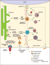
Figure 2. Proposed model for the divergent points of degradative versus secretory autophagy
At the ER exit sites, omegasomes (in mammalian cells) facilitate the formation of degradative autophagosomes, of which the elongation and closure require LC3B. The degradative cargo can be captured/delivered into the degradative autophagosomes via autophagy receptors such as SLRs and TRIMs. Upon closure, degradative autophagosomes display motility toward the minus end of the microtubules where they fuse with lysosomes resulting in the degradation of the engulfed contents by lysosomal hydrolases. Conversely in secretory autophagy, CUPS (in yeast) or their putative (see text for discussion) equivalents in mammalian cells that may be omegasomes located near ER exit sites marked by GRASP localization (as shown for CUPS) aid the formation of secretory autophagosomes. The specific members of mammalian Atg8s that facilitate this process and receptors that capture the cargo into secretory autophagosomes have not been defined. Secretory autophagosomes show vectorial motility toward the plus end of the microtubules and eventually fuse with the plasma membrane releasing their contents into the extracellular environment. The vectorial directionality of secretory autophagic organelles may be controlled by +TIP (plus end tracking proteins). Note that both degradative and secretory autophagosomes may fuse with MVBs to generate amphisomes before fusion with lysosome or plasma membrane, respectively. Degradative and secretory autophagosomes may undergo interconversion or redirection in some instances, e.g. when the same substrate can be secreted or degraded. Either way, the destiny of the contents is removal, whether by lysosomal degradation (degradative autophagy) or plasma membrane extrusion (secretory autophagy). At the bottom left, two recently described processes of autophagy-dependent nonlytic extracellular release of cytoplasmic microbes are depicted: Far left, bacterial ejectosome is represented with the associated autophagosomal profile cup shown to be important for providing membrane to prevent host cell lysis during bacterial ejection. The ejectosome-associated autophagosomal cup is positive for ubiquitin (Ubq), sequestosome 1 (SQSTM1), and Atg8 and its formation requires Atg1, 5, 6, and 7. Left, enterovirus release (nonlytic) is depicted within phosphatidylserine-rich vesicles of autophagic origin that are formed within syntaxin 17-negative, LC3 and Beclin 1-dependent compartments. The examples depicted for autophagic secretion represent two extremes of a potential continuum – from delicate cargo of select proteins or their assemblies to large particulates and whole live microbes.

References

    1. Rabouille C, et al. Diversity in unconventional protein secretion. J Cell Sci. 2012;125:5251–5255. - PubMed
    1. Zhang M, Schekman R. Cell biology. Unconventional secretion, unconventional solutions. Science. 2013;340:559–561. - PubMed
    1. Jiang S, et al. Secretory versus degradative autophagy: unconventional secretion of inflammatory mediators. J Innate Immun. 2013;5:471–479. - PMC - PubMed
    1. Mizushima N, et al. The role of Atg proteins in autophagosome formation. Annu Rev Cell Dev Biol. 2011;27:107–132. - PubMed
    1. Rogov V, et al. Interactions between autophagy receptors and ubiquitin-like proteins form the molecular basis for selective autophagy. Mol Cell. 2014;53:167–178. - PubMed

Publication types