Review
. 2015 Jun:32:162-70.
doi: 10.1016/j.gde.2015.03.006. Epub 2015 May 16.

Mechanisms of myoblast fusion during muscle development

Affiliations
Review

Mechanisms of myoblast fusion during muscle development

Ji Hoon Kim et al. Curr Opin Genet Dev. 2015 Jun.

Abstract

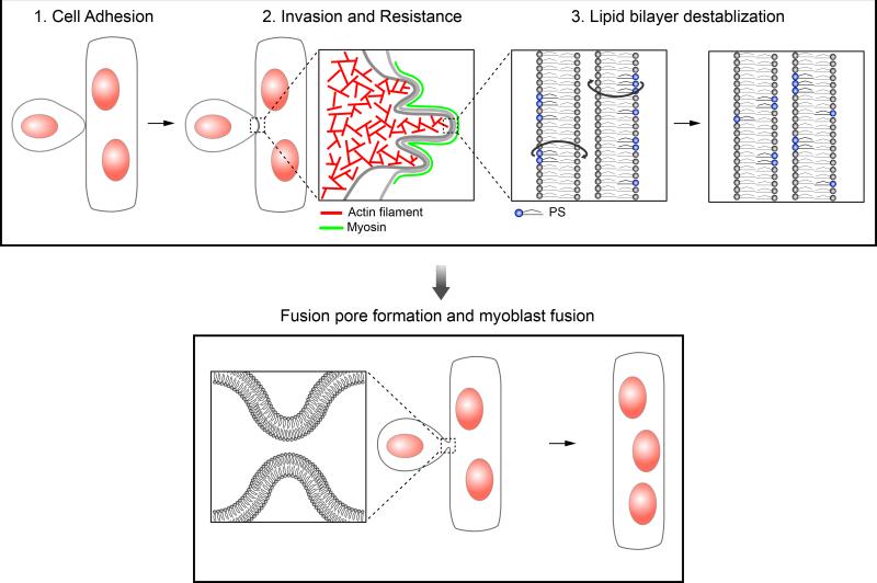
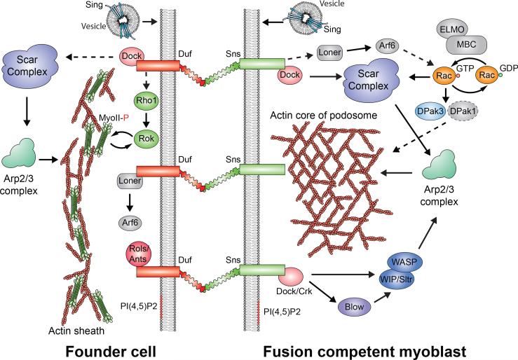
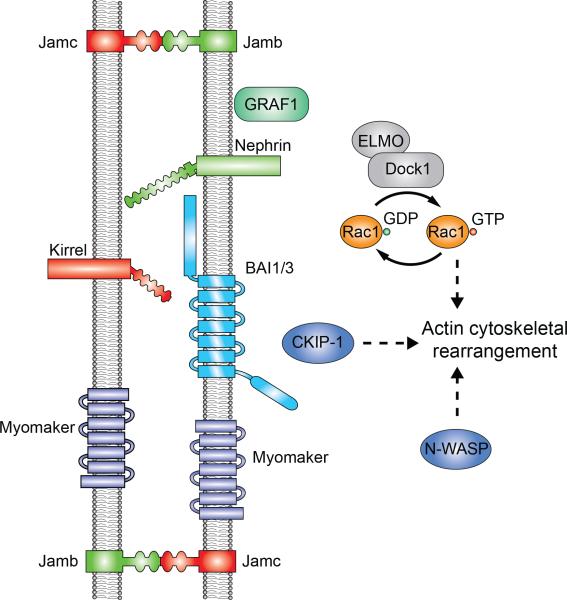
The development and regeneration of skeletal muscle require the fusion of mononucleated muscle cells to form multinucleated, contractile muscle fibers. Studies using a simple genetic model, Drosophila melanogaster, have discovered many evolutionarily conserved fusion-promoting factors in vivo. Recent work in zebrafish and mouse also identified several vertebrate-specific factors required for myoblast fusion. Here, we integrate progress in multiple in vivo systems and highlight conceptual advance in understanding how muscle cell membranes are brought together for fusion. We focus on the molecular machinery at the fusogenic synapse and present a three-step model to describe the molecular and cellular events leading to fusion pore formation.


Figures

Figure 1
Figure 1. The three-step model describing the cellular events leading to fusion pore formation in myoblast fusion
Step 1 – The initial cell adhesion between a founder cell and an FCM is mediated by Ig domain-containing CAMs. Step 2 – Following cell adhesion, the FCM generates actin-propelled membrane protrusions to invade the apposing founder cell, the latter of which mounts a myosin II-mediated mechanosensory response to increase the cortical tension and therefore cortical resistance to the FCM invasion. Step 3 – once the two plasma membranes are brought into close proximity by the invasive and resistance forces, the lipid bilayers are destabilized, leading to the formation of a fusion pore. A hypothetical scenario depicted here is that PS could be flipped from the inner to the outer leaflet, resulting in a more disorganized outer leaflet prone to fusion.
Figure 2
Figure 2. Signaling pathways leading to asymmetric F-actin and myosin accumulation in Drosophila myoblast fusion
Trans-interaction between cell type-specific CAMs, such as Duf (founder cell; with 5 Ig domains) and Sns (FCM; with 8 Ig domains), triggers distinct signaling events in founder cells and FCMs. Briefly, in the founder cell, activation of Scar results in the formation of a thin sheath of F-actin at the fusogenic synapse. In addition, the Rho1-Rok-MyoII pathway amplifies the mechanosensitive accumulation of MyoII at the fusogenic synapse. In the FCM, activation of both Scar and WASP leads to the formation of an F-actin-enriched focus, which is part of an invasive podosome-like structure. For the functions of other components shown here, see the text.
Figure 3
Figure 3. Signaling components required for vertebrate myoblast fusion in vivo
Due to the lack of molecular and cellular markers for the fusogenic synapse in vertebrates, the localization and potential sidedness of these proteins are hypothetical.

References

    1. Buckingham M. Myogenic progenitor cells and skeletal myogenesis in vertebrates. Curr Opin Genet Dev. 2006;16:525–532. - PubMed
    1. Wagers AJ, Conboy IM. Cellular and molecular signatures of muscle regeneration: current concepts and controversies in adult myogenesis. Cell. 2005;122:659–667. - PubMed
    1. Abmayr SM, Pavlath GK. Myoblast fusion: lessons from flies and mice. Development. 2012;139:641–656. [A comprehensive review of components of myoblast fusion uncovered using Drosophila genetics, mouse genetics, and cultured mouse myoblasts.] - PMC - PubMed
    1. Chen EH, Olson EN. Towards a molecular pathway for myoblast fusion in Drosophila. Trends Cell Biol. 2004;14:452–460. - PubMed
    1. Tixier V, Bataille L, Jagla K. Diversification of muscle types: recent insights from Drosophila. Exp Cell Res. 2010;316:3019–3027. - PubMed

Publication types