Review
. 2015 Jul 1;27(25):3717-36.
doi: 10.1002/adma.201501558. Epub 2015 May 19.

Adaptable hydrogel networks with reversible linkages for tissue engineering

Affiliations
Review

Adaptable hydrogel networks with reversible linkages for tissue engineering

Huiyuan Wang et al. Adv Mater. .

Abstract

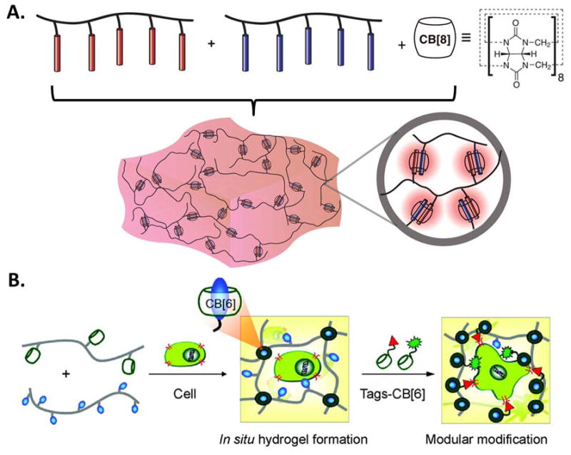
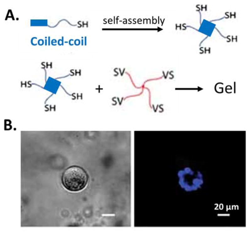
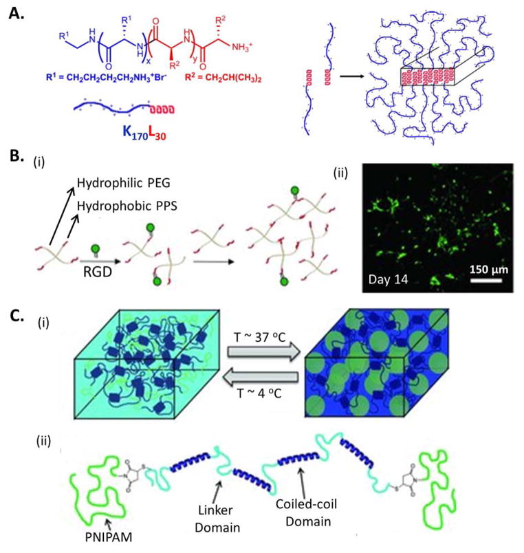
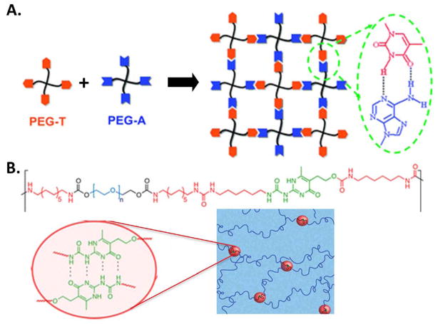
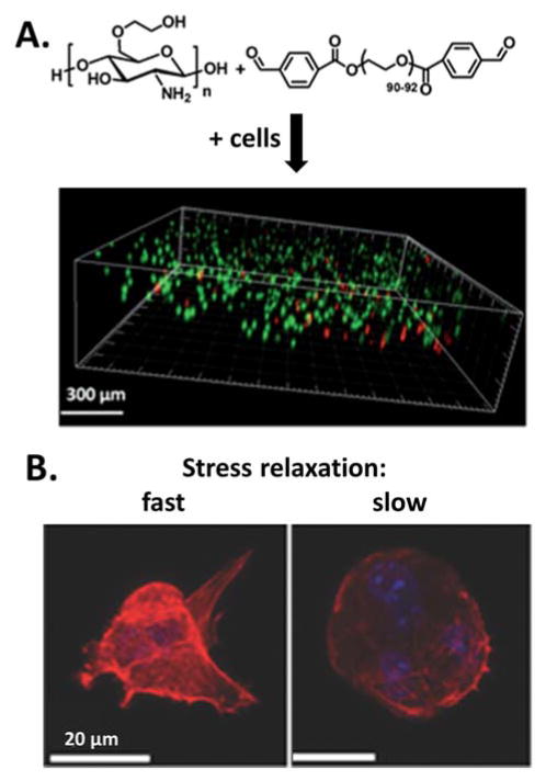
Adaptable hydrogels have recently emerged as a promising platform for three-dimensional (3D) cell encapsulation and culture. In conventional, covalently crosslinked hydrogels, degradation is typically required to allow complex cellular functions to occur, leading to bulk material degradation. In contrast, adaptable hydrogels are formed by reversible crosslinks. Through breaking and re-formation of the reversible linkages, adaptable hydrogels can be locally modified to permit complex cellular functions while maintaining their long-term integrity. In addition, these adaptable materials can have biomimetic viscoelastic properties that make them well suited for several biotechnology and medical applications. In this review, an overview of adaptable-hydrogel design considerations and linkage selections is presented, with a focus on various cell-compatible crosslinking mechanisms that can be exploited to form adaptable hydrogels for tissue engineering.

Keywords: adaptable hydrogels; cell encapsulation; reversible linkages.


Figures

Figure 1
Figure 1
A) Schematic of a permanently crosslinked hydrogel where irreversible degradation occurs. B) Schematic of an adaptable hydrogel built from reversible crosslinks. C) Reversible linkages can be formed either by physical associations or reversible chemical reactions. The dynamics of crosslink breaking and re-forming are related to the kinetic constants of the reactions.
Figure 2
Figure 2
A) Basic design considerations of all hydrogels used as ECM-mimetic materials for cell encapsulation and 3D culture. B) Additional selection criteria for the design of hydrogels with adaptable linkages.
Figure 3
Figure 3
A) Chemical structures, schematic, and physical dimensions of the cyclodextrin (CD) family. Reproduced with permission.[71] Copyright 2012, Royal Society of Chemistry. B) Two guest polymer chains were threaded through γ-CD while only one chain was threaded through β-CD due to differences in CD size. Reproduced with permission.[107] Copyright 2007, American Chemical Society. C) Schematic representation of a shear-thinning hyaluronic acid (HA) hydrogel based on the pendant design of guest-host interactions between adamantine-modified HA and β-cyclodextrin-modified HA. Reproduced with permission.[110] Copyright 2013, American Chemical Society.
Figure 4
Figure 4
A) Schematic of a supramolecular hydrogel prepared through addition of cucurbit[8]uril (CB[8]) to a mixture of two multivalent guest-functionalized polymers. Reproduced with permission.[117] Copyright 2012, American Chemical Society. B) Schematic of a supramolecular hydrogel and its modular modification using host-guest interactions with cucurbit[6]uril (CB[6]) to display bioactive and fluorescent molecules. Reproduced with permission.[119] Copyright 2012, American Chemical Society.
Figure 5
Figure 5
A) Schematic of a mixing-induced, two-component hydrogel formed through biorecognition. Two WW domains (CC43 and a Nedd4.3 variant) bind the same proline-rich peptide (PPxY) with different binding affinities. B) Strain sweeps of C7:P9 and N7:P9 hydrogels demonstrate that tuning of Kd leads to changes in gel stiffness. C) Confocal z-stack projection of neural stem cells differentiated within C7:P9. (red, glial marker, GFAP; green, neuronal marker, MAP2; yellow, progenitor marker, nestin; blue, nuclei, DAPI). Reproduced with permission.[75] Copyright 2009, National Academy of Sciences, U.S.A.
Figure 6
Figure 6
A) Schematic of a hydrogel formed through biorecognition between the PDZ domain in TIP1 protein (CutA-TIP1) and the TIP1-binding peptide. Reproduced with permission.[138] Copyright 2009, Elsevier. B) Proposed structure of the tetrameric ULD-TIP-1 protein (modified from the crystal structures of ULD and TIP-1). Reproduced with permission.[136] Copyright 2012, John Wiley and Sons. C) Schematic of Dock-and-Lock self-assembly mechanism. Docking domains dimerize and lock with the anchoring domains to form shear-thinning hydrogels when mixed. Reproduced with permission.[77] Copyright 2011, Elsevier. D) Schematic of a hydrogel assembled through biorecognition between peptide-binding modules (red) in TPR arrays and peptides coupled to multiarm PEG. Reproduced with permission.[163] Copyright 2012, John Wiley and Sons.
Figure 7
Figure 7
A) Schematic of a hydrogel combining physical crosslinking through the self-assembly of coiled-coil domains and chemical crosslinking between vinyl sulfone (VS) and thiol groups. B) Bright field (left) and fluorescent (right, nucleus staining in blue) of spherical epithelial cell aggregates formed within the hydrogel. Reproduced with permission.[84] Copyright 2011, John Wiley and Sons.
Figure 8
Figure 8
A) Schematic of an amphiphilic diblock copolypeptide hydrogel composed of hydrophilic (blue) and hydrophobic (red) amino acids. Reproduced with permission.[211] Copyright 2009, Elsevier. B) Schematic of an injectable non-protease degradable poly(ethylene glycol)-poly(propylene sulfide) (PEG-PPS) hydrogel formed through hydrophobic interactions of a 4-arm, branched copolymer. Decoration with a cell-adhesive RGD domain enables long-term culture of induced pluripotent stem cell-derived neural progenitor cells (live-dead staining, green: live, red: dead). Reproduced with permission.[213] Copyright 2011, John Wiley and Sons. C) Schematic of a shear-thinning hydrogel formed by coiled-coil self-assembly and reinforced through temperature-responsive aggregation of PNIPAM. Reproduced with permission.[78] Copyright 2013, John Wiley and Sons.
Figure 9
Figure 9
A) Schematic of hydrogel self-assembly via intermolecular, hydrogen bonding of Watson-Crick base pairs between thymine (T) and adenine (A). Reproduced with permission.[233] Copyright 2012, Royal Society of Chemistry. B) Chemical structure of a self-assembling copolymer with a segmented, multiblock architecture including hydrophilic PEG (blue), hydrophobic chain-extenders (red), and UPy groups (green) that undergo self-complementary quadruple H-bonding to form a hydrogel. Reproduced with permission.[238] Copyright 2014, American Chemical Society.
Figure 10
Figure 10
A) Stress relaxation of covalently and ionically crosslinked alginate hydrogels. B) Percentage of cells with stress fibres on elastic and stress-relaxing alginate gels with initial moduli of 1.4_kPa. Data are shown as mean±s.d., and ***P<0.001 (Student’s t-test). C) Representative images of cell spreading on elastic and stress-relaxing alginate gels with initial moduli of 1.4_kPa (actin cytoskeleton in green, paxillin marker for focal adhesions in read). Reproduced with permission.[62] Copyright 2015, Nature Publishing Group.
Figure 11
Figure 11
A) A hydrogel was formed through the dynamic covalent Schiff base linkage between amine groups on chitosan (left structure) and benzaldehyde-modified PEG (right structure) under physiological conditions. Cell viability (live: green, dead: red) and spatial distribution of HeLa cells within the hydrogel after 24 hours is shown. Reproduced with permission.[261] Copyright 2012, Royal Society of Chemistry. B) Representatative images of multicellular behavior within two hydrogels formed through dynamic covalent hydrazone crosslinking. Gels with fast (left) or slow (right) stress relaxation behavior resulted in morphological changes in 3D cell structure after 10 days (F-actin in read and nuclei in blue). Reproduced with permission.[52] Copyright 2013, John Wiley and Sons.

References

    1. Kharkar PM, Kiick KL, Kloxin AM. Chem Soc Rev. 2013;42:7335. - PMC - PubMed
    1. Chan G, Mooney DJ. Trends Biotechnol. 2008;26:382. - PubMed
    1. Cai L, Heilshorn SC. Acta Biomater. 2014;10:1751. - PMC - PubMed
    1. Hoffman BD, Grashoff C, Schwartz MA. Nature. 2011;475:316. - PMC - PubMed
    1. Engler AJ, Sen S, Sweeney HL, Discher DE. Cell. 2006;126:677. - PubMed

Publication types