. 2015 Nov;16(11):2121-33.
doi: 10.1111/pme.12785. Epub 2015 May 19.

The Subjective Experience of Pain: An FMRI Study of Percept-Related Models and Functional Connectivity

Affiliations

The Subjective Experience of Pain: An FMRI Study of Percept-Related Models and Functional Connectivity

Claire E Wilcox et al. Pain Med. 2015 Nov.

Abstract

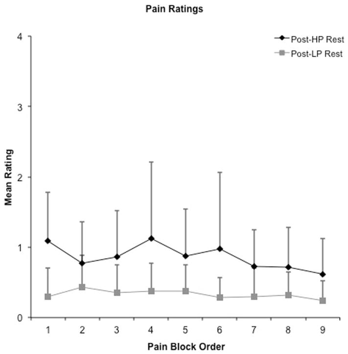
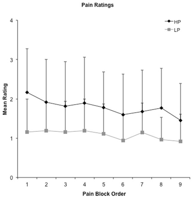
Objective: Previous work suggests that the perception of pain is subjective and dependent on individual differences in physiological, emotional, and cognitive states. Functional magnetic resonance imaging (FMRI) studies have used both stimulus-related (nociceptive properties) and percept-related (subjective experience of pain) models to identify the brain networks associated with pain. Our objective was to identify the network involved in processing subjective pain during cold stimuli.

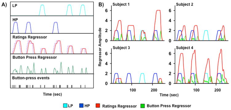
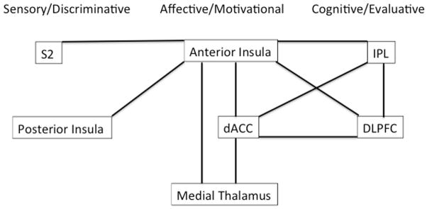
Methods: The current FMRI study directly contrasted a stimulus-related model with a percept-related model during blocks of cold pain stimuli in healthy adults. Specifically, neuronal activation was modelled as a function of changes in stimulus intensity vs as a function of increasing/decreasing levels of subjective pain corresponding to changes in pain ratings. In addition, functional connectivity analyses were conducted to examine intrinsic correlations between three proposed subnetworks (sensory/discriminative, affective/motivational, and cognitive/evaluative) involved in pain processing.

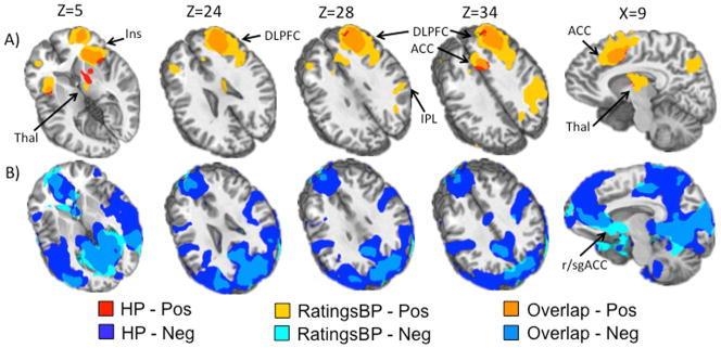
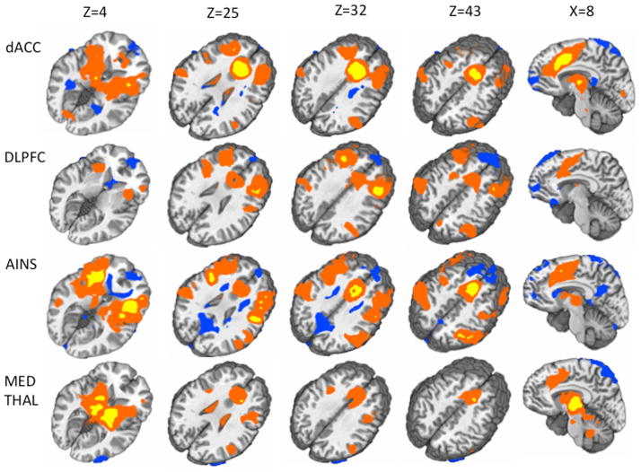
Results: The percept-related model captured more extensive activation than the stimulus-related model and demonstrated an association between higher subjective pain and activation in expected cortical (dorsolateral prefrontal cortex, ventrolateral prefrontal cortex, insula, dorsal anterior cingulate cortex [dACC] extending into pre-supplementary motor area) and subcortical (thalamus, striatum) areas. Moreover, connectivity results supported the posited roles of dACC and insula as key relay sites during neural processing of subjective pain. In particular, anterior insula appeared to link sensory/discriminative regions with regions in the other subnetworks, and dACC appeared to serve as a hub for affective/motivational, cognitive/evaluative, and motor subnetworks.

Conclusions: Using a percept-related model, brain regions involved in the processing of subjective pain during the application of cold stimuli were identified. Connectivity analyses identified linkages between key subnetworks involved in processing subjective pain.

Keywords: Connectivity; Functional Magnetic Resonance Imaging; Pain; Percept-Related; Ratings.


Conflict of interest statement

Disclosures:

The authors have no conflicts of interest to report.

Figures

Figure 1
Figure 1
Panel A depicts sample regressor models. Low (LP; cyan) and high (HP; blue) pain regressors are based on stimulus delivery parameters (onset, duration time). Ratings regressor (red) is modeled on subject’s continuous ratings of pain (purple) convolved with a gamma variate function based on hemodynamic response. Button press regressor (green) is modeled on button press events, convolved with a gamma variate function. Panel B depicts representative graphs for four different subjects plotting the four regressors used in the two hierarchical models. Subjects 1 and 2 demonstrate the expected differentiation in pain ratings (red) between high pain (blue) and low pain (cyan) stimulus blocks, with subject 2 also showing a temporal dissociation between stimulus and ratings onset times. Subject 3 rated all pain stimuli as close to 0 on the Likert scale, while Subject 4 rated all pain stimuli at greater than 5. Button press regressor (green) shows button press event timing.
Figure 2
Figure 2
Mean pain ratings as a function of rest (Top) and pain stimulus (Panel B, gray symbols) blocks. Rest blocks (inter-stimulus intervals) followed all stimulus blocks. Both post high pain block ratings (Post-HP) and high pain ratings (H) (black diamonds) were significantly higher than post low pain block ratings (Post-LP) and low pain ratings (LP) (grey squares) for both rest and pain blocks.
Figure 2
Figure 2
Mean pain ratings as a function of rest (Top) and pain stimulus (Panel B, gray symbols) blocks. Rest blocks (inter-stimulus intervals) followed all stimulus blocks. Both post high pain block ratings (Post-HP) and high pain ratings (H) (black diamonds) were significantly higher than post low pain block ratings (Post-LP) and low pain ratings (LP) (grey squares) for both rest and pain blocks.
Figure 3
Figure 3
This figure presents the results for the high pain regressor and the ratings regressor from the hierarchical regression where regressors were entered in the following order from first to last: low pain, high pain, button press, ratings. Of note the ratings regressor captured more extensive and unique positive activation above and beyond that of the high pain regressor. Panel A (top) shows areas of unique variance associated with the high pain regressor (HP; red), the ratings regressor after correcting for variance associated with the button press regressor (RatingsBP) (yellow), and overlap between the two (orange). Panel B (bottom) shows areas of unique variance associated with HP (dark blue), RatingsBP (light blue) and Overlap (medium blue). Regions indicated by arrows include 1) insula (Ins); 2) thalamus (Thal); 3) anterior cingulate cortex (ACC); 4) dorsolateral prefrontal cortex (DLPFC); 5) inferior parietal lobule (IPL); 6) rostral/subgenual ACC (r/sgACC). Slice locations (Z) are given according to the Talairach atlas.
Figure 4
Figure 4
This figure simplistically depicts the results from the connectivity analyses. It organizes regions into groups according to models which propose central pain-processing networks to be comprised of at least four distinct subnetworks: a sensory/discriminative network, an affective/motivational network, a cognitive evaluative network, and a motor network (not depicted in this figure). Black lines connect regions which were significantly positively correlated with one another (demonstrated connectivity) in our analyses. (S2: secondary somatosensory cortex, dACC: dorsal anterior cingulate cortex, DLPFC: dorsolateral prefrontal cortex, IPL: inferior parietal lobule).
Figure 5
Figure 5
This figure depicts the results from the connectivity analyses. Each row indicates the results for a different seed (from top to bottom: right dACC, right DLPFC, bilateral anterior insula, right medial thalamus). Each column represents a different slice location according to the Talairach atlas (from left to right: Z=4, Z=25, Z=43, X=8). Areas which were significantly positively correlated with seeds (positive Z scores) are displayed in this figure in either yellow (Fischer’s Z>10) or red (Fischer’s Z<10). Areas which were significantly negatively correlated with seeds (negative Z scores) are displayed in blue (Fischer’s Z<10).

References

    1. Apkarian AV, Bushnell MC, Treede RD, Zubieta JK. Human brain mechanisms of pain perception and regulation in health and disease. Eur J Pain. 2005;9:463–84. - PubMed
    1. Wager TD, Atlas LY, Lindquist MA, Roy M, Woo CW, Kross E. An fMRI-based neurologic signature of physical pain. N Engl J Med. 2013;368:1388–97. - PMC - PubMed
    1. Davis KD, Moayedi M. Central mechanisms of pain revealed through functional and structural MRI. J Neuroimmune Pharmacol. 2012;8:518–34. - PubMed
    1. Duerden EG, Albanese MC. Localization of pain-related brain activation: a meta-analysis of neuroimaging data. Hum Brain Mapp. 2013;34:109–49. - PMC - PubMed
    1. Porro CA, Baraldi P, Pagnoni G, Serafini M, Facchin P, Maieron M, et al. Does anticipation of pain affect cortical nociceptive systems? J Neurosci. 2002;22:3206–14. - PMC - PubMed

Publication types