Review
. 2015 Sep;11(3):277-305.
doi: 10.1007/s11302-015-9452-9. Epub 2015 May 20.

Purinergic signalling during development and ageing

Affiliations
Review

Purinergic signalling during development and ageing

Geoffrey Burnstock et al. Purinergic Signal. 2015 Sep.

Abstract

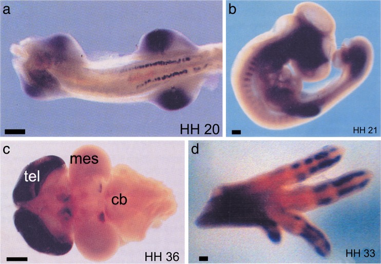
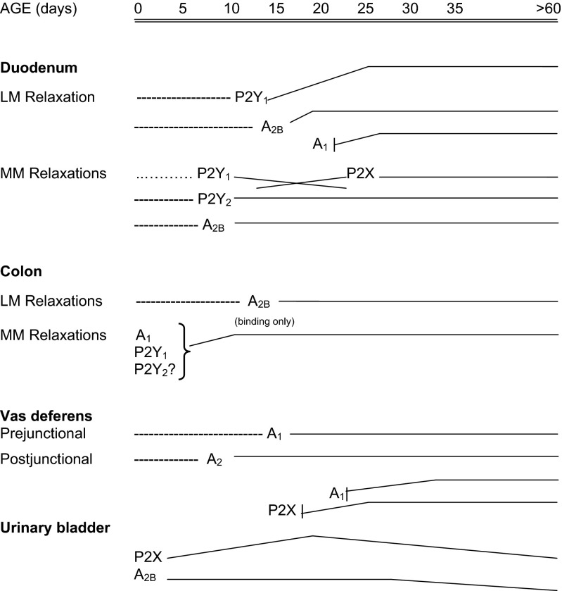
Extracellular purines and pyrimidines play major roles during embryogenesis, organogenesis, postnatal development and ageing in vertebrates, including humans. Pluripotent stem cells can differentiate into three primary germ layers of the embryo but may also be involved in plasticity and repair of the adult brain. These cells express the molecular components necessary for purinergic signalling, and their developmental fates can be manipulated via this signalling pathway. Functional P1, P2Y and P2X receptor subtypes and ectonucleotidases are involved in the development of different organ systems, including heart, blood vessels, skeletal muscle, urinary bladder, central and peripheral neurons, retina, inner ear, gut, lung and vas deferens. The importance of purinergic signalling in the ageing process is suggested by changes in expression of A1 and A2 receptors in old rat brains and reduction of P2X receptor expression in ageing mouse brain. By contrast, in the periphery, increases in expression of P2X3 and P2X4 receptors are seen in bladder and pancreas.


Figures

Fig. 1
Fig. 1
Purinergic signalling is upstream of the eye field transcription factors (EFTFs) that are expressed very early in development of the frog embryo and to control development of the eye (see [364]). ATP released in the presumptive eye field is converted to ADP via NTPDase2. The ADP can activate the P2Y1 receptor (P2Y1R), which then triggers expression of the transcription factor Rx1
Fig. 2
Fig. 2
Morpholino oligonucleotide knockdown of NTPDase2 (NTPD2MO) or the P2Y1 receptor (P2Y1MO) alters the expression of Pax6 and Rx1 (a–h). The morpholino constructs were injected on just one side of the early embryo to facilitate a comparison with the uninjected side as an internal control. NTPD2MO either on its own or in combination with P2Y1MO reduced Pax6 and Rx1 expression only on the injected side (arrows). However, injection of P2Y1MO on its own or a control morpholino (CMO) has no effect on expression of Pax6 and Rx1 (ik). This reduced expression of Pax6 and Rx1 results in smaller eyes on the injected side (indicated by lacZ staining) or, in the case of the dual morpholino knockdown, no eyes (k, arrow) (reproduced with permission from [26])
Fig. 3
Fig. 3
The expression of the components of purinergic signalling and Pax6 and Otx2 during frog embryonic development. a NTPDase2 expression precedes induction of Pax6, and expression of the P2Y1R is coincident with the upregulation of Pax6 that occurs at St 12.5. b Both NTPDase2 and P2Y1 are expressed more strongly in the mesoderm/endoderm, suggesting that it is this tissue layer that initiates expression of the EFTFs in the ectoderm. c ATP is released from the neural plate during development. The inset diagram indicates the positioning of ATP biosensors in the very early frog embryo. A large transient signal is seen in the anterior region—the site of the eye field. ATP signalling is also seen in the posterior regions. The ATP release event is blown up in the inset traces (reproduced with permission from [26])
Fig. 4
Fig. 4
Interaction between acetylcholine (ACh) and ATP recorded in an otocyst from chick embryo. a The response to 10 μM ACh. b The response to 100 μM ATP. c The response to the coapplication of 10 μM ACh and 100 μM ATP. The records in ac were taken in this order at 5-min intervals. The bath solutions contained 25 mM Ca2+ (reproduced with permission from [30])
Fig. 5
Fig. 5
Expression of P2Y1 receptors during embryonic development of the chick as visualized by whole-mount in situ hybridization. Stages of development are shown in bottom right corner. a Ventral view of stage 20 embryo showing P2Y1 expression in mesonephros and limb buds (scale bar = 200 μm). b Lateral view of the chick somite at stage 21 showing P2Y1 expression in the anterior region. The dark area in the head region is due to an artefact of photography (scale bar = 200 μm). c Dorsal view of stage 36 brain (anterior to the left), showing increased levels of expression in telecephlon (tel), dorsal diencephlon and posterior midbrain. mes mesencephalon, cb cerebellum (scale bar = 1 mm). d An anterior-uppermost view of a leg at embryonic stage 33. Expression of P2Y1 is seen in the digits, but not in areas of joint formation. The same expression pattern is also seen in the wing (scale bar = 100 μm) (reproduced with permission from [34])
Fig. 6
Fig. 6
K+ responses to ATP analogues of a mouse mesodermal cell line. Each of the two traces was obtained from the same cell (ae). The responses induced by ATP (left traces) and ATP analogues (right traces) are shown. The names of the analogues are shown near the traces. Each drug was applied at 20 μM, and the holding potentials were 0 mV (reproduced with permission from [39])
Fig. 7
Fig. 7
Diagram summarizing the development of functional responses mediated by purine receptors in the rat duodenum, colon, urinary bladder and vas deferens. The dashed lines represent ages at which it was not possible to study functional responses, and the solid lines show when responses were observed, with the slope of the line indicating whether a response, in general, increased or decreased with age (reproduced with permission from [264])

References

    1. Burnstock G. Purine and pyrimidine receptors. Cell Mol Life Sci. 2007;64:1471–1483. - PMC - PubMed
    1. Yeung CH. Temporary inhibition of the initiation of motility of demembranated hamster sperm by high concentrations of ATP. Int J Androl. 1986;9:359–370. - PubMed
    1. Foresta C, Rossato M, Di Virgilio F. Extracellular ATP is a trigger for the acrosome reaction in human spermatozoa. J Biol Chem. 1992;267:1–5. - PubMed
    1. Kupitz Y, Atlas D. A putative ATP-activated Na+ channel involved in sperm-induced fertilization. Science. 1993;261:484–486. - PubMed
    1. Smith R, Koenig C, Pereda J. Adenosinetriphosphatase (Mg-ATPase) activity in the plasma membrane of preimplantation mouse embryo as revealed by electron microscopy. Anat Embryol (Berl) 1983;168:455–466. - PubMed

LinkOut - more resources