Case Reports
. 2015 Aug;35(4):603-9.
doi: 10.1007/s10792-015-0081-7. Epub 2015 May 20.

Aflibercept versus ranibizumab for treating persistent diabetic macular oedema

Affiliations
Case Reports

Aflibercept versus ranibizumab for treating persistent diabetic macular oedema

Kristof R O A Vandekerckhove. Int Ophthalmol. 2015 Aug.

Abstract

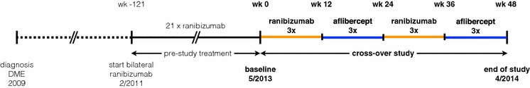
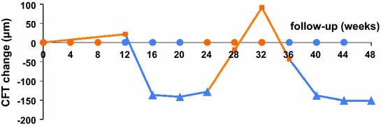
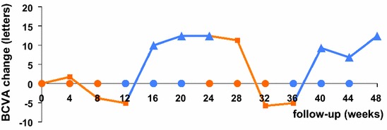
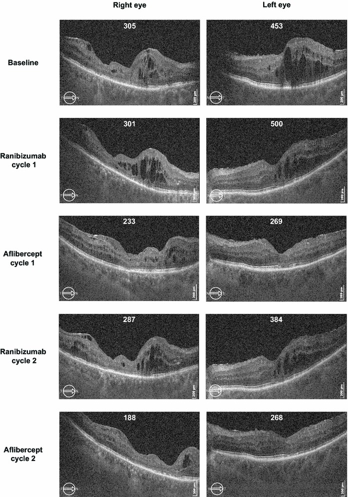
The purpose of this study was to compare the efficacy of intravitreal aflibercept versus ranibizumab for treating therapy-resistant diabetic macular oedema (DME). A 69-year-old man presented with persistent bilateral DME despite previous ranibizumab treatment. Bilateral study treatment comprised one cycle of three monthly ranibizumab injections (0.5 mg), followed by one cycle of three aflibercept injections (2.0 mg), a second ranibizumab cycle and a second aflibercept cycle. Baseline visual acuity (ETDRS score) was 60 letters for the right eye and 65 letters for the left eye. Baseline central foveal thickness (CFT) was 305 μm for the right eye and 453 μm for the left eye. Substantially improved outcomes were observed during the first aflibercept cycle. CFT was reduced by 150 μm (mean) in both the eyes and decreased below the lowest level achieved during the previous 2.5-year ranibizumab treatment. Visual acuity was improved by 17.5 letters (mean) in both the eyes. Reintroduction of ranibizumab immediately worsened the status of both eyes back to the baseline level. During the final aflibercept cycle, visual acuity and CFT improved to the same levels achieved during the first aflibercept cycle. In this case study, we prospectively switched the treatment three times and observed a dramatic and consistent treatment advantage for aflibercept.


Figures

Fig. 1
Fig. 1
Flowchart describing the study design (one patient, both the eyes). Study injections and examinations were performed at 4-week intervals (±2 days). wk week
Fig. 2
Fig. 2
Graph showing changes in mean CFT from baseline over 48 weeks, during treatment with ranibizumab or aflibercept. The mean for both the eyes is shown. Note the consistent response during the ranibizumab and aflibercept treatment cycles. Orange squares response 4 weeks after ranibizumab, blue triangles response 4 weeks after aflibercept, orange dots ranibizumab injections, blue dots aflibercept injections, CFT Central Foveal Thickness
Fig. 3
Fig. 3
Graph showing changes in mean BCVA (ETDRS letters) from baseline over 48 weeks, during treatment with ranibizumab or aflibercept. The mean for both the eyes is shown. Note the consistent response during the ranibizumab and aflibercept treatment cycles. Orange squares response 4 weeks after ranibizumab, blue triangles response 4 weeks after aflibercept, orange dots ranibizumab injections, blue dots aflibercept injections, BCVA best-corrected visual acuity, ETDRS Early Treatment of Diabetic Retinopathy Study
Fig. 4
Fig. 4
Successive ocular coherence tomography (OCT) of horizontal sections (7.0 mm) from the right and left eyes shows the evolution of subretinal and intraretinal fluid. Central foveal thickness (μm) is indicated for each OCT exam. Images correspond to the OCT scans taken 4 weeks after the last (third) injection of each treatment cycle. Baseline image was taken after a 1-month washout period after the prestudy treatment (near-monthly ranibizumab injections). Clear improvement is observed during the two aflibercept treatment cycles, whereas worsening is observed during the two ranibizumab treatment cycles. CFT Central Foveal Thickness

Similar articles

Cited by

References

    1. Nguyen QD, Brown DM, Marcus DM, et al. Ranibizumab for diabetic macular edema: results from 2 phase III randomized trials: RISE and RIDE. Ophthalmology. 2012;119:789–801. doi: 10.1016/j.ophtha.2011.12.039. - DOI - PubMed
    1. Korobelnik JF, Do DV, Schmidt-Erfurth U, et al. Intravitreal aflibercept for diabetic macular edema. Ophthalmology. 2014;121:2247–2254. doi: 10.1016/j.ophtha.2014.05.006. - DOI - PubMed
    1. Gregori NZ, Feuer W, Rosenfeld PJ. Novel method for analyzing snellen visual acuity measurements. Retina. 2010;30:1046–1050. doi: 10.1097/IAE.0b013e3181d87e04. - DOI - PubMed
    1. Papadopoulos N, Martin J, Ruan Q, et al. Binding and neutralization of vascular endothelial growth factor (VEGF) and related ligands by VEGF Trap, ranibizumab and bevacizumab. Angiogenesis. 2012;15:171–185. doi: 10.1007/s10456-011-9249-6. - DOI - PMC - PubMed
    1. Muether PS, Droege KM, Fauser S. Vascular endothelial growth factor suppression times in patients with diabetic macular oedema treated with ranibizumab. Br J Ophthalmol. 2014;98:179–181. - PubMed

Publication types

MeSH terms

LinkOut - more resources