. 2015 Jul;104(1):225-34.e3.
doi: 10.1016/j.fertnstert.2015.04.021. Epub 2015 May 16.

Stro-1/CD44 as putative human myometrial and fibroid stem cell markers

Affiliations

Stro-1/CD44 as putative human myometrial and fibroid stem cell markers

Aymara Mas et al. Fertil Steril. 2015 Jul.

Abstract

Objective: To identify and characterize myometrial/fibroid stem cells by specific stem cell markers in human myometrium, and to better understand the stem cell contribution in the development of uterine fibroids.

Design: Prospective, experimental human and animal study.

Setting: University research laboratory.

Patient(s)/animal(s): Women undergoing hysterectomy for treatment of symptomatic uterine fibroids and female NOD/SCID/IL-2Rγ(null) mice.

Intervention(s): Identification and isolation of stem cells from human fibroids and adjacent myometrium tissues using Stro-1/CD44-specific surface markers.

Main outcome measure(s): Flow cytometry, semiquantitative polymerase chain reaction, clonogenicity assays, cell culture, molecular analysis, immunocyto-histochemistry, in vitro differentiation, and xenotransplantation assays.

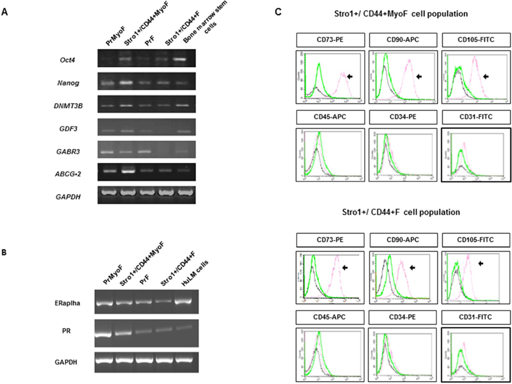
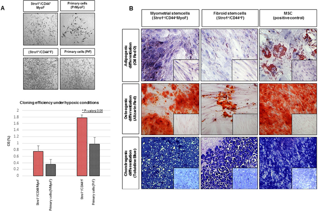
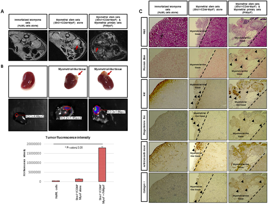
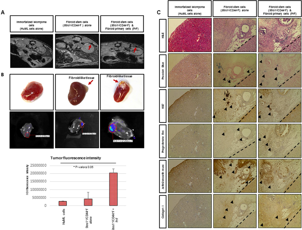
Result(s): Using Stro-1/CD44 surface markers, we were able to isolate stem cells from adjacent myometrium and human fibroid tissues. The undifferentiated status of isolated cells was confirmed by the expression of ABCG2 transporter, as well as additional stem cell markers OCT4, NANOG, and GDB3, and the low expression of steroid receptors ERα and PR-A/PR-B. Mesodermal cell origin was established by the presence of typical mesenchymal markers (CD90, CD105, and CD73) and absence of hematopoietic stem cell markers (CD34, CD45), and confirmed by the ability of these cells to differentiate in vitro into adipocytes, osteocytes, and chondrocytes. Finally, their functional capability to form fibroid-like lesions was established in a xenotransplantation mouse model. The injected cells labeled with superparamagnetic iron oxide were tracked by both magnetic resonance imaging and fluorescence imaging, thus demonstrating the regenerative potential of putative fibroid stem cells in vivo.

Conclusion(s): We have demonstrated that Stro-1/CD44 can be used as specific surface markers to enrich a subpopulation of myometrial/fibroids cells, exhibiting key features of stem/progenitor cells. These findings offer a useful tool to better understand the initiation of uterine fibroids, and may lead to the establishment of effective therapeutic options.

Keywords: CD44/Stro-1; Human myometrium; stem cells; uterine fibroids/leiomyomas.


Figures

Figure 1
Figure 1. Molecular characterization of isolated MyoF and F stem and primary cells
(A) PCR assays demonstrating the presence and/or absence of undifferentiated genes in Stro-1+/CD44+MyoF/F stem cells versus primary cells PrMyoF/PrF (B) Molecular characterization showing the mRNA expression of hormonal receptors (ERalpha,PR) in primary cells PrMyoF/PrF versus Stro-1+/CD44+MyoF/F stem cells (C) Representative flow cytometric analysis of Stro-1+/CD44+MyoF/F displaying the positive expression of CD73, CD90, CD105. A negative expression of Stro-1+/CD44+MyoF/F was observed for hematopoietic stem cell markers (CD45 and CD34), endothelial marker (CD31).
Figure 2
Figure 2. In vitro studies: Clonogenic assay and multipotential differentiation
(A) Highlighted macroscopic morphology showing the colonies formed by Stro-1+/CD44+Myo F (upper left), Stro-1+/CD44+F (upper right) and matching primary cells PrMyoF/PrF (lower left and right) cultured at 156 cell/cm2 for 15 days under hypoxic conditions. Lower graph shows cloning efficiency (%) of Stro-1+/CD44+MyoF and Stro-1+/CD44+F stem versus primary cells PrMyoF/PrF (data are means ± SEM). (B) (Upper pannel) Induction of adipogenic differentiation detected by the presence of Oil Red-O staining in Stro-1+/CD44+Myo F and Stro-1+/CD44+F cells. (Middle pannel) Osteogenic differentiation of the induced Stro-1+/CD44+MyoF and Stro-1+/CD44+F cells identified by the specific Alizarin Red staining and (Lower pannel) induction of chondrocyte differentiation as detected by staining with Toluidine Blue. MSC cells were included as a positive control with typical Oil Red-O, Alizarin red and Toluidine Blue staining. Untreated Stro-1+/CD44+MyoF and Stro-1+/CD44+F cells were incorporated as negative controls (see small pictures). All histological images were examined under a 20X objective lens.
Figure 3
Figure 3. In vivo reconstruction of human myometrial/fibroid tissues in kidney capsule of NOD-SCID mice injected with MyoF cells
(A) In vivo xenograft imaging of mice using a magnetic resonance labeling (MRI) scanner. Scans were performed the week before to animal sacrifice and seven weeks after the injection of super-paramagnetic iron oxide (SPIO)-labeled Stro-1+/CD44+MyoF cells. HuML cells were used as a negative control. The recruitment of SPIO-labeled Stro-1+/CD44+MyoF cells resulted in decrease of signal intensity (SI) and the visualization of darker areas in xenograft sites. (B) Upper panels revealed the xenograft generated in the kidney capsule at macroscopic level. Middle panels represent In vivo xenograft imaging of mice using using the IVIS (Xenogen, Lincolnshire, United Kingdom) for 120 seconds as well the. The images obtained displayed a color spectrum, with the weakest signal being in blue and the strongest red. This intensity of signal is represented in the lower graph. (C) Upper panels shows the H&E staining, demonstrating the characteristic histology of fibroid-like tissue containing some adipose cells and Prussian Blue dye, allowing us to localize the human cells due to the accumulation of iron deposits. We can also appreciate the Ki67 staining, demonstrating the proliferative ability of these cells. Lower panels revealed the human Progesterone Receptor signal in the nucleus of xenograft cells from fibroid-like tissue, suggesting the role of P4 in the growth and maintenance of leiomyoma reconstructed tissues and SMA staining, specific of smooth muscle cells. Finally, we showed the COL1A1 staining that validates the enrichment of extracellular matrix in the xenograft formed. All images were captured with a 10-fold magnification.
Figure 4
Figure 4. In vivo reconstruction of human myometrial/fibroid tissues in kidney capsule of NOD-SCID mice injected with F cells
(A) In vivo xenograft imaging of mice using a magnetic resonance labeling (MRI) scanner. Scans were performed a week prior to animal sacrifice and seven weeks after the injection of super-paramagnetic iron oxide (SPIO)-labeled Stro-1+/CD44+F cells. HuML cells were used as a negative control. The recruitment of SPIO-labeled Stro-1+/CD44+F cells resulted in decrease of signal intensity (SI) and the visualization of darker areas in xenograft sites. (B) Upper panels revealed the xenograft generated in the kidney capsule at macroscopic level. Middle panels represent In vivo xenograft imaging of mice using using the IVIS (Xenogen, Lincolnshire, United Kingdom) for 120 seconds as well the. The images obtained displayed a color spectrum, with the weakest signal being in blue and the strongest red. This intensity of signal is represented in the lower graph. (C) Upper panels shows the H&E staining, demonstrating the characteristic histology of fibroid-like tissue containing some adipose cells and Prussian Blue dye, allowing us to localize the human cells due to the accumulation of iron deposits. We can also appreciate the Ki67 staining, demonstrating the proliferative ability of these cells. Lower panels revealed the human Progesterone Receptor signal in the nucleus of xenograft cells from fibroid-like tissue, suggesting the role of P4 in the growth and maintenance of leiomyoma reconstructed tissues and SMA staining, specific of smooth muscle cells. Finally, we showed the COL1A1 staining that validates the enrichment of extracellular matrix in the xenograft formed. All images were captured with a 10fold magnification.

References

    1. Zimmermann A, Bernuit D, Gerlinger C, Schaefers M, Geppert K. Prevalence, symptoms and management of uterine fibroids: an international internet-based survey of 21,746 women. BMC Womens Health. 2012;12:6. - PMC - PubMed
    1. Stewart EA. Uterine fibroids. Lancet. 2001;357:293–298. - PubMed
    1. Parker WH. Uterine myomas: management. Fertil Steril. 2007;88(2):255–271. Review. - PubMed
    1. Mas A, Cervello I, Gil-Sanchis C, Simón C. Current understanding of somatic stem cells in leiomyoma formation. Fertil Steril. 2014;102(3):613–620. - PubMed
    1. Cardozo ER, Clark AD, Banks NK, Henne MB, Stegmann BJ, Segars JH. The estimated annual cost of uterine leiomyomata in the United States. Am J Obstet Gynecol. 2012;206:211. e1–9. - PMC - PubMed

Publication types

MeSH terms