. 2015 May 20:5:10433.
doi: 10.1038/srep10433.

Cyclin-dependent kinase 11(p110) (CDK11(p110)) is crucial for human breast cancer cell proliferation and growth

Affiliations

Cyclin-dependent kinase 11(p110) (CDK11(p110)) is crucial for human breast cancer cell proliferation and growth

Yubing Zhou et al. Sci Rep. .

Abstract

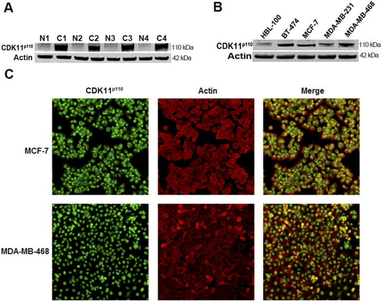
Cyclin-dependent kinases (CDKs) play important roles in the development of many types of cancers by binding with their paired cyclins. However, the function of CDK11 larger protein isomer, CDK11(p110), in the tumorigenesis of human breast cancer remains unclear. In the present study, we explored the effects and molecular mechanisms of CDK11(p110) in the proliferation and growth of breast cancer cells by determining the expression of CDK11(p110) in breast tumor tissues and examining the phenotypic changes of breast cancer cells after CDK11(p110) knockdown. We found that CDK11(p110) was highly expressed in breast tumor tissues and cell lines. Tissue microarray analysis showed that elevated CDK11(p110) expression in breast cancer tissues significantly correlated with poor differentiation, and was also associated with advanced TNM stage and poor clinical prognosis for breast cancer patients. In vitro knockdown of CDK11(p110) by siRNA significantly inhibited cell growth and migration, and dramatically induced apoptosis in breast cancer cells. Flow cytometry demonstrated that cells were markedly arrested in G1 phase of the cell cycle after CDK11(p110) downregulation. These findings suggest that CDK11(p110) is critical for the proliferation and growth of breast cancer cells, which highlights CDK11(p110) may be a promising therapeutic target for the treatment of breast cancer.


Conflict of interest statement

The authors declare no competing financial interests.

Figures

Figure 1
Figure 1
CDK11p110 is highly expressed in human breast cancer tissues and cell lines. (A) Breast tumor tissues and their adjacent normal tissues were lysed and immunoblotted to determine CDK11p110 expression, with Actin as an internal reference. All the gels were run under the same experimental conditions. Representative example of CDK11p110 expression in breast cancer tissues (C1-C4) and adjacent normal tissues (N1-N4) are shown. (B) Levels of CDK11p110 expression in normal human breast cell line HBL-100 and four breast cancer cell lines BT-474, MCF-7, MDA-MB-231, and MDA-MB-468 were detected using Western blot. All the gels were run under the same experimental conditions. (C) Expression of CDK11p110 in MCF-7 and MDA-MB-468 cells was assessed by immunofluorescence with antibodies to CDK11p110 and Actin. Cells were visualized under a fluorescence microscope after incubation with secondary fluorescent conjugated antibodies Alexa Fluor 488 goat anti-rabbit IgG (green) or Alexa Fluor 594 goat anti-mouse IgG (red).
Figure 2
Figure 2
CDK11p110 expression levels correlate with clinicopathological characteristics of breast cancer patients. CDK11p110 levels in breast tumor tissue microarray were determined by immunohistochemistry, and the correlation of CDK11p110 expression with clinicopathological characteristics of breast cancer patients was evaluated. (A) Distribution of CDK11p110 staining scores among breast tumor tissues and their adjacent normal tissues. (B) Distribution of CDK11p110 staining scores among histological grade II and grade III breast tumor tissues. (C) Distribution of CDK11p110 staining scores among TNM II stage and III stage breast tumor tissues. (D) Distribution of CDK11p110 staining scores among survivor and nonsurvivor breast tumor tissues. (E) Kaplan-Meier survival curve of breast cancer patients with CDK11p110 low staining (CDK11p110 staining ≤3) or high staining (CDK11p110 staining ≥4). (F) Representative images of different immunohistochemical staining intensities of CDK11p110 in breast cancer tissues. On the basis of the percentage of cells with positive nuclear staining, CDK11p110 staining patterns were categorized into 6 groups: 0, no nuclear staining; 1+: < 10% positive cells; 2+, 10%-25% positive cells; 3+, 26%-50% positive cells; 4+, 51%-75% positive cells; 5+, > 75% positive cells. Original magnification ×400.
Figure 3
Figure 3
CDK11p110 knockdown inhibits human breast cancer cell proliferation in vitro. Human CDK11p110 siRNA were transfected into MCF-7 and MDA-MB-468 cells, followed by cell proliferation determination. (A and B) Morphological changes of MCF-7 and MDA-MB-468 cells, respectively, after CDK11p110 knockdown for 72 hours. (C and D) Cell viability changes determined by MTT assay of MCF-7 and MDA-MB-468 cells, respectively, after CDK11p110 knockdown for 72 hours. *P < 0.05, **P < 0.01 compared with the 0 nM siRNA group. (E and F) Downregulation of CDK11p110 mRNA and protein expression, determined by RT-PCR and Western blot, respectively, by CDK11p110 siRNA in MCF-7 and MDA-MB-468 cells. All the gels were run under the same experimental conditions. (1: 0 nM siRNA, 2: 40 nM nonspecific siRNA, 3: 10 nM CDK11p110 siRNA, 4: 20 nM CDK11p110 siRNA, 5: 40 nM CDK11p110 siRNA). (G and H) CDK11p110 knockdown induces cell death and decreases CDK11p110 expression in MCF-7 and MDA-MB-468 cells, respectively, detected by immunofluorescence. (I) Macroscopic images of cell colony formation alterations of MCF-7 and MDA-MB-468 cells, after CDK11p110 knockdown with 40 nM of CDK11p110 siRNA. (J and K) Cell colony formation rate changes of MCF-7 and MDA-MB-468 cells, respectively, after CDK11p110 knockdown with 40 nM of CDK11p110 siRNA. **P < 0.01 compared with the cell only group.
Figure 4
Figure 4
CDK11p110 knockdown suppresses human breast cancer cell migration. After transfection with 20 nM of CDK11p110 siRNA or nonspecific siRNA for 72 hours, the cell migration activity of MCF-7 and MDA-MB-468 cells was assessed by the wound healing assay. (A and B) Morphological images of MCF-7 and MDA-MB-468 cell migration, respectively, after CDK11p110 knockdown. (C and D) Migration distance of MCF-7 and MDA-MB-468 cells, respectively, after CDK11p110 knockdown. **P < 0.01 compared with the cell only group.
Figure 5
Figure 5
CDK11p110 knockdown induces cell apoptosis in human breast cancer cells. After transfection with 20 nM of CDK11p110 siRNA or nonspecific siRNA for 72 hours, the cell apoptosis of MCF-7 and MDA-MB-468 cells was assessed by flow cytometry analysis and Western blot. (A and B) Representative images of cell apoptosis alterations of MCF-7 and MDA-MB-468 cells, respectively, after CDK11p110 knockdown for 72 hours. (C and D) Apoptosis rate of MCF-7 and MDA-MB-468 cells, respectively, after CDK11p110 knockdown. **P < 0.01 compared with the cell only group. (E and F) Representative images of apoptosis-related proteins alterations in MCF-7 and MDA-MB-468 cells, respectively, after CDK11p110 knockdown for 72 hours. All the gels were run under the same experimental conditions. (1: 0 nM siRNA, 2: 40 nM nonspecific siRNA, 3: 10 nM CDK11p110 siRNA, 4: 20 nM CDK11p110 siRNA, 5: 40 nM CDK11p110 siRNA).
Figure 6
Figure 6
CDK11p110 knockdown induces human breast cancer cell cycle arrest in G1 phase. After transfection with 20 nM of CDK11p110 siRNA or nonspecific siRNA for 72 hours, the cell cycle distribution of MCF-7 and MDA-MB-468 cells was determined by flow cytometry. (A and B) Representative images of cell cycle distribution alterations of MCF-7 and MDA-MB-468 cells, respectively, after CDK11p110 knockdown. (C and D) Cell number in different cell cycle phases of MCF-7 and MDA-MB-468 cells, respectively, after CDK11p110 knockdown. *P < 0.05, **P < 0.01 compared with the cell only group.

References

    1. Torre L. A. et al. Global cancer statistics, 2012. CA. Cancer J. Clin. 65, 87–108 (2015). - PubMed
    1. DeSantis C., Ma J., Bryan L. & Jemal A. Breast cancer statistics, 2013. CA. Cancer J. Clin. 64, 52–62 (2014). - PubMed
    1. Shah R., Rosso K. & Nathanson S. D. Pathogenesis, prevention, diagnosis and treatment of breast cancer. World J. Clin. oncol. 5, 283–298 (2014). - PMC - PubMed
    1. Hosford S. R. & Miller T. W. Clinical potential of novel therapeutic targets in breast cancer: CDK4/6, Src, JAK/STAT, PARP, HDAC, and PI3K/AKT/mTOR pathways. Pharmgenomics Pers. Med. 7, 203–215 (2014). - PMC - PubMed
    1. Mohamed A., Krajewski K., Cakar B. & Ma C. X. Targeted therapy for breast cancer. Am. J. Pathol. 183, 1096–1112 (2013). - PubMed

Publication types

Substances