. 2015 Aug;14(4):635-43.
doi: 10.1111/acel.12346. Epub 2015 May 20.

Lgr5⁺ amacrine cells possess regenerative potential in the retina of adult mice

Affiliations

Lgr5⁺ amacrine cells possess regenerative potential in the retina of adult mice

Mengfei Chen et al. Aging Cell. 2015 Aug.

Abstract

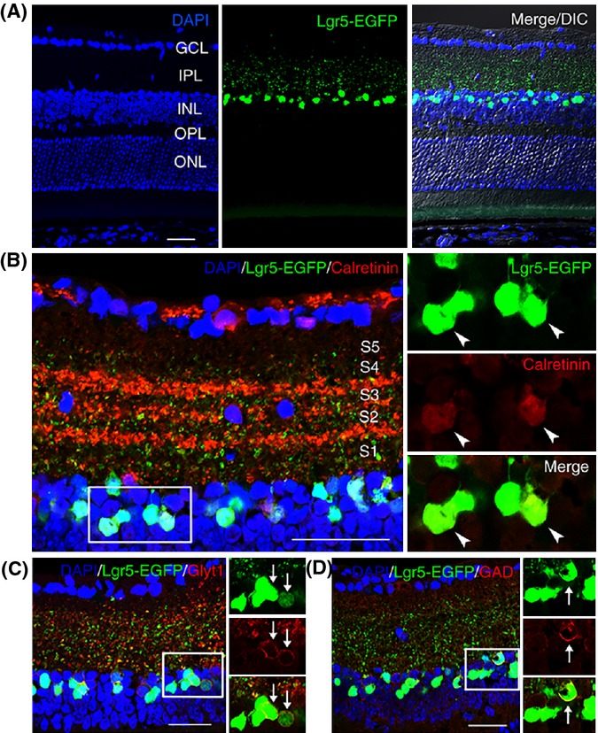
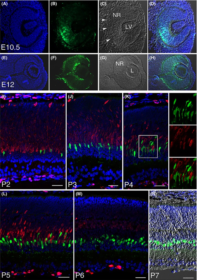
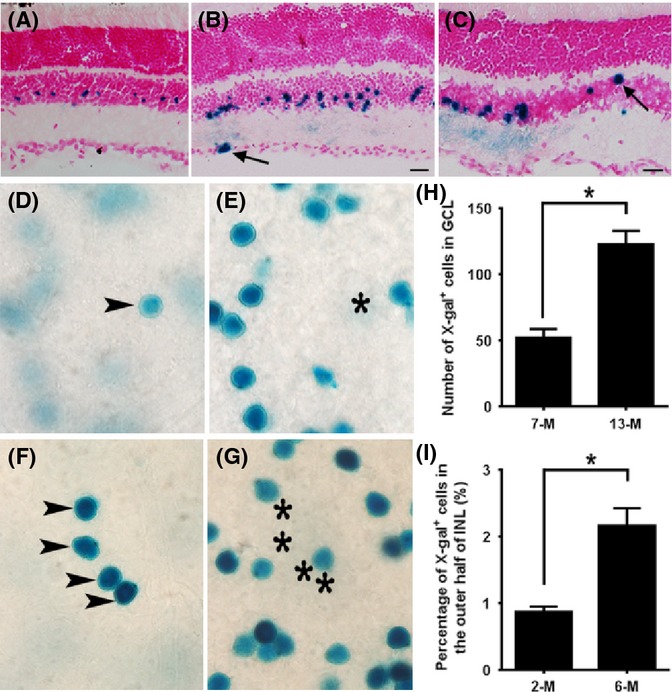
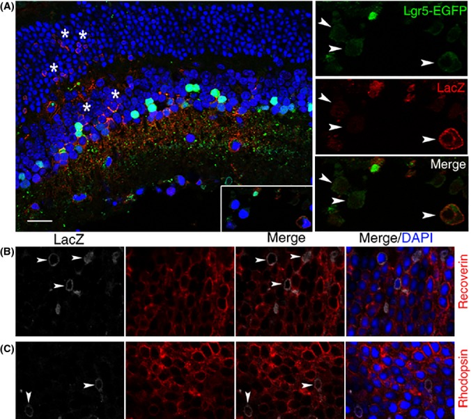
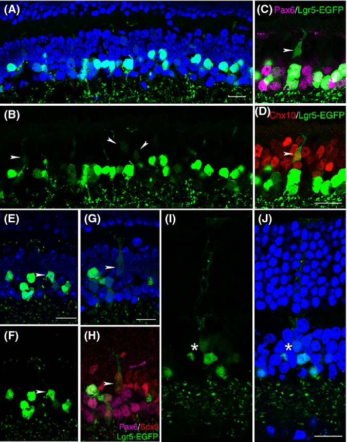
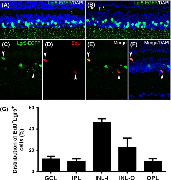
Current knowledge indicates that the adult mammalian retina lacks regenerative capacity. Here, we show that the adult stem cell marker, leucine-rich repeat-containing G-protein-coupled receptor 5 (Lgr5), is expressed in the retina of adult mice. Lgr5(+) cells are generated at late stages of retinal development and exhibit properties of differentiated amacrine interneurons (amacrine cells). Nevertheless, Lgr5(+) amacrine cells contribute to regeneration of new retinal cells in the adult stage. The generation of new retinal cells, including retinal neurons and Müller glia from Lgr5(+) amacrine cells, begins in early adulthood and continues as the animal ages. Together, these findings suggest that the mammalian retina is not devoid of regeneration as previously thought. It is rather dynamic, and Lgr5(+) amacrine cells function as an endogenous regenerative source. The identification of such cells in the mammalian retina may provide new insights into neuronal regeneration and point to therapeutic opportunities for age-related retinal degenerative diseases.

Keywords: Lgr5; aging; amacrine cells; neurogenesis; retina; retinal regeneration.


Figures

Fig 1
Fig 1
Lgr5-EGFP marks a subset of amacrine cells in the inner nuclear layer of adult mouse retina. (A) Confocal images of EGFP expression from the Lgr5 locus (Lgr5-EGFP) in the retina of 8-week-old Lgr5EGFP-Ires-CreERT2 mice. Lgr5-EGFP+ cells are restricted to the inner nuclear layer with axons projecting to the inner plexiform layer. (B) Staining of Lgr5EGFP-Ires-CreERT2 mouse retina with anti-calretinin antibody. The majority of Lgr5-EGFP+ cells are also positive for calretinin. The processes of Lgr5-EGFP+ cells can reach sublaminal layer 5 (S5) within the inner plexiform layer. (C) Staining of Lgr5EGFP-Ires-CreERT2 mouse retina with anti-GlyT1 antibody. (D) Staining of Lgr5EGFP-Ires-CreERT2 mouse retina with anti-GAD antibody. Boxed areas in panels (B–D) are highlighted in adjacent higher magnification views, with overlapping signals indicated by arrows or arrowheads. GCL, ganglion cell layer; IPL, inner plexiform layer; INL, inner nuclear layer; OPL, outer plexiform layer; ONL, outer nuclear layer. Scale bars = 30 μm.
Fig 2
Fig 2
Lgr5-EGFP expression at various stages of retinogenesis. (A–D) Lgr5-EGFP expression in the developing eye is first detected in the optic cup at E10.5, beginning in the middle of the optic cup and then expanding to its peripheral margin. Arrowheads in C highlight retinal pigmented epithelium. NR, neural retina; LV, lens vesicle. (E–H) Lgr5-EGFP expression in the neural retina (NR), lens (L), retinal pigmented epithelium, and surrounding mesenchymal cells at E12. (I–M) Confocal images of Lgr5EGFP-Ires-CreERT2 mouse retina co-stained with Ki67 (red) at postnatal day 2 through day 6. Lgr5-EGFP+ cells do not express Ki67 at any stage of late retinogenesis, indicating that they have exited the cell cycle. Boxed area in panel K is highlighted in the adjacent panels allowing for higher magnification views. (N) Merged image of DAPI staining, Lgr5-EGFP, and DIC of a retinal section at P7. In all relevant panels, Lgr5-EGFP is in green and DAPI staining is in blue. Scale bars = 30 μm.
Fig 3
Fig 3
Lineage tracing of Lgr5-EGFP+ cells in mouse retinas. (A–C) X-gal staining (blue) of retinas from Lgr5EGFP-Ires-CreERT2; Rosa26-LacZ mice at 3 weeks (A), 2 months (B), and 6 months (C) of age following tamoxifen injection at P3 to P4 (A, B) or 6 weeks of age (C). Arrows mark LacZ-positive cells in the retinal ganglion layer (B) and outer edge of the inner nuclear layer (C). (D–G) Images of whole-mount retinal samples stained with X-gal following tamoxifen injection at 4–6 weeks of age. Images in panels D and E were taken from the same location of the same retina sample, with D focused to the ganglion cell layer and E focused to the inner nuclear layer. The X-gal+ cell located in the ganglion cell layer (D) is highlighted with an arrowhead. Its corresponding vertical location in the inner nuclear layer was marked with a star (E). Similar to panels D and E, X-gal staining images in panels F and G are taken from the same retinal sample at the same location, with panel F focused to the ganglion cell layer and panel G focused to the inner nuclear layer. At this location, four X-gal+ cells form a cluster in the ganglion cell layer. (H) Numbers of X-gal+ cells in the ganglion cell layer in 7-month-old and 13-month-old mice. n = 6 retinas from three mice of each group. *P = 0.004 by Student’s t-test. (I) Percentage of X-gal-positive cells that are located in the outer half of the inner nuclear layer in 2-month-old and 6-month-old mice. n = 8 and 12 sections from three mice of each group. *P = 0.01 by Student’s t-test. Scale bars = 20 μm.
Fig 4
Fig 4
Generation of new retinal cells in response to retinal injury. (A) Confocal images of a retinal cross section from Lgr5EGFP-Ires-CreERT2; Rosa26-LacZ mice analyzed with anti-β-galactosidase (LacZ) antibody (red) following retinal NMDA and growth factor injection. LacZ-positive cells are present in all three cell layers of the retina (indicated by stars in the inner nuclear layer and outer nuclear layer). The adjacent higher magnification views show co-localization of the Lgr5-EGFP signal with LacZ in cells of the loosely organized retinal ganglion layer (highlighted with arrowheads). (B) Co-staining of LacZ with recoverin (a marker for photoreceptors). Arrowhead marks LacZ and recoverin double-positive photoreceptor cells. (C) Co-staining of LacZ with rhodopsin (a marker for rod photoreceptors). Arrowheads mark LacZ and rhodopsin double-positive photoreceptor cells. Scale bars = 20 μm.
Fig 5
Fig 5
Generation of retinal neurons and Müller glial cells by Lgr5-EGFP+ amacrine cells in the retina under normal physiological conditions. (A, B) Confocal images of strong and weak Lgr5-EGFP-expressing cells in the inner nuclear layer of 6-week-old Lgr5EGFP-Ires-CreERT2 mice. Weakly Lgr5-EGFP-expressing cells are marked with arrowheads. (C, D) Dual immunohistochemical analysis of Lgr5-EGFP with Pax6 (C) and Lgr5-EGFP with Chx10 (D). In these views, the arrowhead marks a weak Lgr5-EGFP expression immature bipolar cell that lost Pax6 expression and was positive for Chx10 staining. (E, F) A Lgr5-EGFP+ cell with typical mature bipolar cell morphology, highlighted with arrowheads. (G, H) A weak Lgr5-EGFP-expressing immature Müller cell. In triple stainings of panel H, the weak Lgr5-EGFP-expressing immature Müller cell was negative for Pax6 staining, but positive for Sox9 staining. (I, J) A weakly Lgr5-EGFP-expressing mature Müller cell in the retina of a 15-month-old mouse. The star marks the cell body of the weakly Lgr5-EGFP-expressing cell. Nuclei are stained by DAPI (blue) in panels A, E, G, and J. Scale bars = 20 μm.
Fig 6
Fig 6
Labeling of Lgr5-EGFP+ cells with EdU in the retina of adult mice. (A, B) Confocal images of the retina obtained from a 2-month-old Lgr5EGFP-Ires-CreERT2 mouse (A) and an 8-month-old Lgr5EGFP-Ires-CreERT2 mouse (B). Stars in panel B highlight the processes of some Lgr5-EGFP+ cells that project into the outer plexiform layer at this age. (C–F) Confocal images of the retina from a 2.5-month-old Lgr5EGFP-Ires-CreERT2 mouse labeled with EdU. EdU and Lgr5-EGFP double-positive cells are highlighted with arrowheads. (G) Distribution of EdU and Lgr5-EGFP double-positive cells across the retina. n = 62 sections from three mice. GCL, ganglion cell layer; IPL, inner plexiform layer; INL-I, inner half of inner nuclear layer; INL-O, outer half of the inner nuclear layer; OPL, outer plexiform layer; ONL, outer nuclear layer.

References

    1. Ahmad I, Tang L, Pham H. Identification of neural progenitors in the adult mammalian eye. Biochem. Biophys. Res. Commun. 2000;270:517–521. - PubMed
    1. Amato MA, Arnault E, Perron M. Retinal stem cells in vertebrates: parallels and divergences. Int. J. Dev. Biol. 2004;48:993–1001. - PubMed
    1. Barker N, van Es JH, Kuipers J, Kujala P, van den Born M, Cozijnsen M, Haegebarth A, Korving J, Begthel H, Peters PJ, Clevers H. Identification of stem cells in small intestine and colon by marker gene Lgr5. Nature. 2007;449:1003–1007. - PubMed
    1. Bernardos RL, Barthel LK, Meyers JR, Raymond PA. Late-stage neuronal progenitors in the retina are radial Muller glia that function as retinal stem cells. J. Neurosci. 2007;27:7028–7040. - PMC - PubMed
    1. Bhatia B, Singhal S, Jayaram H, Khaw PT, Limb GA. Adult retinal stem cells revisited. Open Ophthalmol. J. 2010;4:30–38. - PMC - PubMed

Publication types

MeSH terms

Substances