

High resolution pituitary gland MRI at 7.0 tesla: a clinical evaluation in Cushing's disease
- PMID: 25991481
- PMCID: PMC4666272
- DOI: 10.1007/s00330-015-3809-x
High resolution pituitary gland MRI at 7.0 tesla: a clinical evaluation in Cushing's disease
Abstract
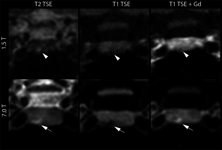
Objective: To evaluate the detection of pituitary lesions at 7.0 T compared to 1.5 T MRI in 16 patients with clinically and biochemically proven Cushing's disease.
Methods: In seven patients, no lesion was detected on the initial 1.5 T MRI, and in nine patients it was uncertain whether there was a lesion. Firstly, two readers assessed both 1.5 T and 7.0 T MRI examinations unpaired in a random order for the presence of lesions. Consensus reading with a third neuroradiologist was used to define final lesions in all MRIs. Secondly, surgical outcome was evaluated. A comparison was made between the lesions visualized with MRI and the lesions found during surgery in 9/16 patients.
Results: The interobserver agreement for lesion detection was good at 1.5 T MRI (κ = 0.69) and 7.0 T MRI (κ = 0.62). In five patients, both the 1.5 T and 7.0 T MRI enabled visualization of a lesion on the correct side of the pituitary gland. In three patients, 7.0 T MRI detected a lesion on the correct side of the pituitary gland, while no lesion was visible at 1.5 T MRI.
Conclusion: The interobserver agreement of image assessment for 7.0 T MRI in patients with Cushing's disease was good, and lesions were detected more accurately with 7.0 T MRI.
Key points: Interobserver agreement for lesion detection on 1.5 T MRI was good; Interobserver agreement for lesion detection on 7.0 T MRI was good; 7.0 T enabled confirmation of unclear lesions at 1.5 T; 7.0 T enabled visualization of lesions not visible at 1.5 T.
Keywords: Cushing’s disease, pituitary; Magnetic resonance imaging; Pituitary ACTH hypersecretion; Pituitary adenoma; Pituitary gland.
Figures

References
-
- Juszczak A, Grossman A. The management of Cushing's disease - from investigation to treatment. Endokrynol Pol. 2013;64:166–174. - PubMed
-
- Newell-Price J, Trainer P, Besser M, Grossman A. The diagnosis and differential diagnosis of Cushing's syndrome and pseudo-Cushing's states. Endocr Rev. 1998;19:647–672. - PubMed
-
- Witek P, Zielinski G. Predictive value of preoperative magnetic resonance imaging of the pituitary for surgical cure in Cushing's disease. Turk Neurosurg. 2012;22:747–752. - PubMed
Publication types
MeSH terms
LinkOut - more resources
Full Text Sources
Other Literature Sources
Medical
