. 2015 Jul 17;290(29):17669-17678.
doi: 10.1074/jbc.M114.626275. Epub 2015 May 19.

Defective Guanine Nucleotide Exchange in the Elongation Factor-like 1 (EFL1) GTPase by Mutations in the Shwachman-Diamond Syndrome Protein

Affiliations

Defective Guanine Nucleotide Exchange in the Elongation Factor-like 1 (EFL1) GTPase by Mutations in the Shwachman-Diamond Syndrome Protein

Adrián García-Márquez et al. J Biol Chem. .

Abstract

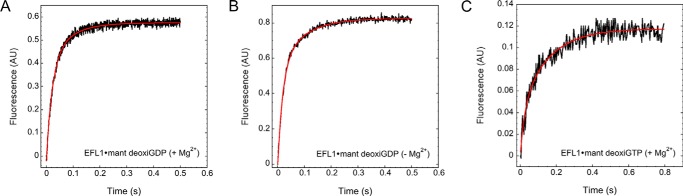
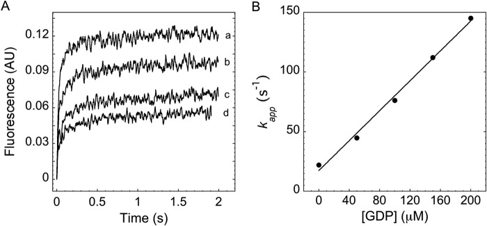
Ribosome biogenesis is orchestrated by the action of several accessory factors that provide time and directionality to the process. One such accessory factor is the GTPase EFL1 involved in the cytoplasmic maturation of the ribosomal 60S subunit. EFL1 and SBDS, the protein mutated in the Shwachman-Diamond syndrome (SBDS), release the anti-association factor eIF6 from the surface of the ribosomal subunit 60S. Here we report a kinetic analysis of fluorescent guanine nucleotides binding to EFL1 alone and in the presence of SBDS using fluorescence stopped-flow spectroscopy. Binding kinetics of EFL1 to both GDP and GTP suggests a two-step mechanism with an initial binding event followed by a conformational change of the complex. Furthermore, the same behavior was observed in the presence of the SBDS protein irrespective of the guanine nucleotide evaluated. The affinity of EFL1 for GTP is 10-fold lower than that calculated for GDP. Association of EFL1 to SBDS did not modify the affinity for GTP but dramatically decreased that for GDP by increasing the dissociation rate of the nucleotide. Thus, SBDS acts as a guanine nucleotide exchange factor (GEF) for EFL1 promoting its activation by the release of GDP. Finally, fluorescence anisotropy measurements showed that the S143L mutation present in the Shwachman-Diamond syndrome altered a surface epitope for EFL1 and largely decreased the affinity for it. These results suggest that loss of interaction between these proteins due to mutations in the disease consequently prevents the nucleotide exchange regulation the SBDS exerts on EFL1.

Keywords: EFL1; GTPase; Shwachman-Diamond syndrome protein; fluorescence anisotropy; fluorescence resonance energy transfer (FRET); guanine nucleotide exchange factor (GEF); guanine nucleotides; ribosome assembly.


Figures

FIGURE 1.
FIGURE 1.
Binding of mant-GDP to EFL1 alone or in complex with SBDS. Mant fluorescence was excited by FRET from the intrinsic tryptophan residues in EFL1. Concentrations after mixing consisted of 4 μm EFL1, 10 μm SBDS, 50 μm mant-GDP, and 5 mm Mg2+ as relevant for the corresponding experiment. Gray traces correspond to the fit to a double exponential equation. A, association of mant-GDP to EFL1. B, dissociation of mant-GDP from EFL1 with 500 μm GDP. C, concentration dependence of kapp values for mant-GDP association to EFL1; circles, kapp,1; squares, kapp,2. D, association of mant-GDP to EFL1 in the absence of Mg2+. E, dissociation of mant-GDP from EFL1 with 500 μm GDP in the absence of Mg2+. F, concentration dependence of kapp values for mant-GDP association to EFL1 in the absence of Mg2+; circles, kapp,1; squares, kapp,2. G, association of mant-GDP to the EFL1·SBDS complex. H, dissociation of mant-GDP from the EFL1·SBDS complex with 500 μm GDP. I, concentration dependence of kapp values for mant-GDP association to the EFL1·SBDS complex; circles, kapp,1; squares, kapp,2.
FIGURE 2.
FIGURE 2.
Binding of mant-GTP to EFL1 alone or in complex with SBDS. Mant fluorescence was excited by FRET from the intrinsic tryptophan residues in EFL1. Concentrations after mixing consisted of 4 μm EFL1, 10 μm SBDS, 50 μm mant-GTP/Gpp(NH)p, and 5 mm Mg2+ as relevant for the corresponding experiment. Gray traces correspond to the fit to a double exponential equation. A, association of mant-GTP to EFL1. B, concentration dependence of kapp values for mant-GTP association to EFL1; circles, kapp,1; squares, kapp,2. C, association of mant-GTP to EFL1 in the absence of Mg2+. D, concentration dependence of kapp values for mant-GTP association to EFL1 in the absence of Mg2+; circles, kapp,1; squares, kapp,2. E, association of mant-GTP to the EFL1·SBDS complex. F, concentration dependence of kapp values for mant-GTP association to the EFL1·SBDS complex; circles, kapp,1; squares, kapp,2. G, association of mant-Gpp(NH)p to EFL1. H, concentration dependence of kapp values for mant-Gpp(NH)p association to EFL1; circles, kapp,1; squares, kapp,2.
FIGURE 3.
FIGURE 3.
Binding kinetics of the EFL1 protein produced in bacteria to mant-GDP. A, fit to a single exponential model and B, fit to a double exponential model.
FIGURE 4.
FIGURE 4.
Binding of mant-deoxyguanine nucleotides to EFL1. Mant fluorescence was excited by FRET from the intrinsic tryptophan residues in EFL1. Concentrations after mixing consisted of 4 μm EFL1, 50 μm mant-deoxy-GDP/deoxy-GTP, and 5 mm Mg2+ as relevant for the corresponding experiment. Gray traces correspond to the fit to a double exponential equation. A, association of mant-deoxy-GDP to EFL1. B, association of mant-deoxy-GDP to EFL1 in the absence of Mg2+. C, association of mant-deoxy-GTP to EFL1.
FIGURE 5.
FIGURE 5.
Competition binding kinetics of EFL1 to mant-GDP and unmodified GDP. A, association of mant-GDP to EFL1 in the presence of increasing concentrations of unmodified GDP: 0 (a), 50 (b), 150 (c), and 200 (d) μm GDP. B, concentration dependence of the kapp value for GDP association to EFL1.
FIGURE 6.
FIGURE 6.
Characterization of the interaction between human EFL1 and human SBDS wild-type and disease mutant S143L. A, fluorescence anisotropy measurement of the interaction between SBDS-FlAsH wild-type (black circles) and disease mutation SBDS-FlAsH S143L (gray circles) with EFL1. B, secondary structure analysis of SBDS wild-type (black) and disease mutation SBDS S143L (gray) evaluated by far UV-CD.
FIGURE 7.
FIGURE 7.
Kinetic scheme of the interaction of EFL1 with guanine nucleotides. A, in the presence of 5 mm Mg2+ and B, in the absence of Mg2+.

References

    1. Kressler D., Hurt E., Bassler J. (2010) Driving ribosome assembly. Biochim. Biophys. Acta 1803, 673–683 - PubMed
    1. Thomson E., Ferreira-Cerca S., Hurt E. (2013) Eukaryotic ribosome biogenesis at a glance. J. Cell Sci. 126, 4815–4821 - PubMed
    1. Woolford J. L., Jr., Baserga S. J. (2013) Ribosome biogenesis in the yeast Saccharomyces cerevisiae. Genetics 195, 643–681 - PMC - PubMed
    1. McCann K. L., Baserga S. J. (2013) Genetics: mysterious ribosomopathies. Science 341, 849–850 - PMC - PubMed
    1. Teng T., Thomas G., Mercer C. A. (2013) Growth control and ribosomopathies. Curr. Opin. Genet. Dev. 23, 63–71 - PubMed

Publication types

MeSH terms

LinkOut - more resources