. 2015 Apr;19(2):141-50.
doi: 10.1055/s-0034-1396522. Epub 2014 Dec 8.

Lipidomic profiling of mastoid bone and tissue from patients with chronic otomastoiditis

Affiliations

Lipidomic profiling of mastoid bone and tissue from patients with chronic otomastoiditis

Farbod Fazlollahi et al. Int Arch Otorhinolaryngol. 2015 Apr.

Abstract

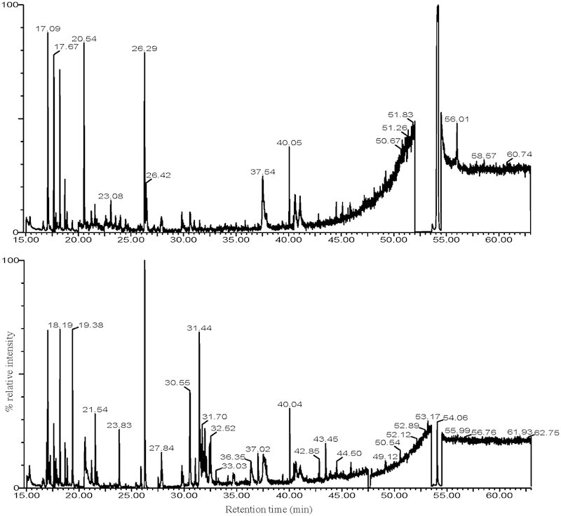
Introduction Chronic otomastoiditis causes pain, otorrhea, and hearing loss resulting from the growth of tissue within the normally hollow mastoid cavity. Objectives In this report, we used a lipidomics approach to profile major mastoid bone and tissue lipids from patients with and without otomastoiditis. Methods The bone dust created during mastoidectomy, as well as the mastoid tissue, was analyzed from seven patients. Bone dust was also collected and analyzed in an additional four otologic cases (parotidectomy requiring mastoidectomy). Samples were subjected to a modified Bligh/Dyer lipid extraction, then high-performance thin-layer chromatography (HPTLC), combined gas chromatography/electron impact-mass spectrometry (GC/EI-MS), and flow-injection/electrospray ionization-tandem mass spectrometry (FI/ESI-MSMS). Data were analyzed for identification and profiling of major lipid components. Results HPTLC revealed the presence of various lipid classes, including phosphatidylcholines, cholesterol, and triacylglycerols. GC/EI-MS analysis revealed the presence of cholesterol and several fatty acids. FI/ESI-MSMS analysis revealed a host of phosphatidylcholines, phosphatidylethanolamines, and cholesteryl esters. Conclusion We used a lipidomics approach to develop an efficient (both in time and tissue amount) methodology for analysis of these tissues, identify the most abundant and common lipid species, and create a base of knowledge from which more focused endeavors in biomarker discovery can emerge. In an effort toward improved patient categorization and individualized intervention, the ultimate goal of this work is to correlate these lipid molecules to disease state and progression. This is the first reported study of its kind on these tissues.

Keywords: flow injection analysis; lipid metabolism; mass spectrometry; mastoiditis.


Figures

Fig. 1
Fig. 1
Orcinol-stained (top panel) and Coomassie blue-stained (bottom panel) high-performance thin-layer chromatography (HPTLC) displays of (left to right): 1, standards (5 μg/compound except CerLac 2 μg); 2, mastoid tissue; 4, bone dust; 6, blank. Matched blood controls for each sample are in the adjoining lanes (for example, for lane 2 the matched blood control is in lane 3). The plates were run and first stained with orcinol and then Coomassie as described in the Methods. The HPTLC-purified samples that were separately prepared for mass spectrometric analysis were: zone 1, Rf 0.0 to 0.24; zone 2 Rf 0.24 to 0.47; zone 3 Rf 0.47 to 0.71; zone 4 Rf 0.71 to 1.0. Abbreviations: C, cholesterol; CerLac, lactosylceramide; CerPCho, sphingomyelin; TAG, triacylglycerol; PtdCho, phosphatidylcholine.
Fig. 2
Fig. 2
Combined gas chromatography/electron impact-mass spectrometry total ion current chromatograms from high-performance thin-layer chromatography (HPTLC) zone 4 extracts. Total ion current chromatograms of HPTLC zone 4 trimethylsilyl (TMS)-derivatized eluates from bone dust (top panel) collected during mastoidectomy and tissue excised from the mastoid cavity (bottom panel) in patients with chronic otomastoiditis. The dominant peak in each chromatogram (retention time 54.06 minutes) was identified as cholesterol. Other tentative identifications include: glycerol (20.54 minutes), palmitic acid (37.02 minutes), oleic acid (40.59 minutes), and stearic acid (41.04 minutes). The region from 15.00 to 20.00 minutes is populated by byproducts of the derivatization reaction and excess derivatizing reagents, and the large peak at 26.25 minutes in the bottom panel was identified at butylated hydroxytoluene. To aid in visualizing smaller peaks, the ordinate of each chromatogram was regionally expanded as follows: top panel: 15.03 to 17.71 (×10), 17.79 to 19.89 (×24), 19.99 to 52.00 (×186), 54.50 to 63.00 (×54); bottom panel: 15.03 to 17.51 (×16), 17.69 to 25.99 (×36), 27.49 to 47.50 (×54), 47.70 to 53.50 (×36), 54.50 to 63.00 (×24). The fatty acids observed in these chromatograms were present in abundance below the limit of confident identification, thus the spectra were not of high quality.
Fig. 3
Fig. 3
Representative tandem mass spectrometry spectra of the majorly present lipid classes in mastoid tissue. (a) Precursor ion scan of m/z 369.4 shows cholesteryl esters and similar compounds. (b) Precursor ion scan of m/z 184 of samples shows phosphatidylcholines, sphingomyelins, and similar compounds. (c) Neutral loss scan of m/z 141 of samples shows phosphatidylethanolamines and similar compounds. (d) Precursor ion scan of m/z 264.1 shows ceramides and similar compounds. Compounds not shown here were present below the limit of detection.

Similar articles

References

    1. Rovers M M, Schilder A G, Zielhuis G A, Rosenfeld R M. Otitis media. Lancet. 2004;363(9407):465–473. - PubMed
    1. Morris P S Richmond P Lehmann D Leach A J Gunasekera H Coates H L New horizons: otitis media research in Australia Med J Aust 2009191(9, Suppl):S73–S77. - PubMed
    1. Ameer F, Scandiuzzi L, Hasnain S, Kalbacher H, Zaidi N. De novo lipogenesis in health and disease. Metabolism. 2014;63(7):895–902. - PubMed
    1. de Kroon A I, Rijken P J, De Smet C H. Checks and balances in membrane phospholipid class and acyl chain homeostasis, the yeast perspective. Prog Lipid Res. 2013;52(4):374–394. - PubMed
    1. Luthra A, Denisov I G, Sligar S G. Spectroscopic features of cytochrome P450 reaction intermediates. Arch Biochem Biophys. 2011;507(1):26–35. - PMC - PubMed