Leiomyomatosis peritonealis disseminata positive for progesterone receptor
- PMID: 25992687
- PMCID: PMC4444146
- DOI: 10.12659/AJCR.893570
Leiomyomatosis peritonealis disseminata positive for progesterone receptor
Abstract
Background: Leiomyomatosis peritonealis disseminata (LPD) is a rare condition that occurs in reproductive-age women. The pathogenesis of LPD is considered to be related to female sex hormones.
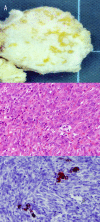
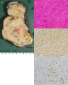
Case report: A 30-year-old woman who had undergone an ovariectomy due to calcified thecoma at 24 years of age and had delivered a baby boy at 29 years of age showed abnormal abdominal-pelvic masses in a computed tomography scan. The peritoneal nodules were resected and histologically diagnosed as LPD. Smooth muscle cells in LPD lesions expressed progesterone receptor, while estrogen receptor and luteinizing hormone/chorionic gonadotropin receptor were negative.
Conclusions: LPD should be considered when multiple nodules mimicking dissemination of malignancies are found in the abdominal cavity. In the present case, progesterone may have been involved in the pathogenesis of LPD.
Figures


References
-
- Willson JR, Peale AR. Multiple peritoneal leiomyomas associated with a granulosa-cell tumor of the ovary. Am J Obstet Gynecol. 1952;64:204–8. - PubMed
-
- Al-Talib A, Tulandi T. Pathophysiology and possible iatrogenic cause of leiomyomatosis peritonealis disseminata. Gynecol Obstet Invest. 2010;69:239–44. - PubMed
-
- Kouakou F, Gondo RAD, N’Guessan VLK, et al. Leiomyomatosis peritonealis disseminata and pregnancy: a case report. Clin Exp Obstet Gynecol. 2012;39:252–54. - PubMed
-
- Barone A, Ambrosio MR, Rocca BJ, et al. Leiomyomatosis peritonealis disseminata: an additional case. Eur J Gynaecol Oncol. 2014;35:188–91. - PubMed
Publication types
MeSH terms
Substances
LinkOut - more resources
Full Text Sources
Medical
Research Materials

