. 2015 May 20;10(5):e0127102.
doi: 10.1371/journal.pone.0127102. eCollection 2015.

A pilot study to assess effects of long-term inhalation of airborne particulate matter on early Alzheimer-like changes in the mouse brain

Affiliations

A pilot study to assess effects of long-term inhalation of airborne particulate matter on early Alzheimer-like changes in the mouse brain

Dhaval P Bhatt et al. PLoS One. .

Abstract

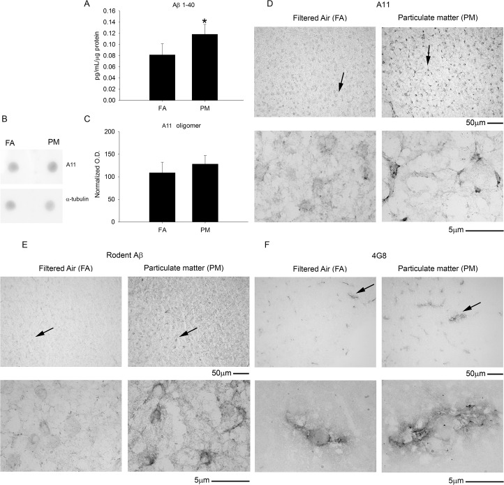
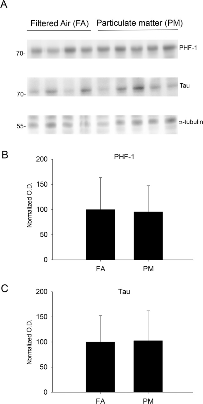
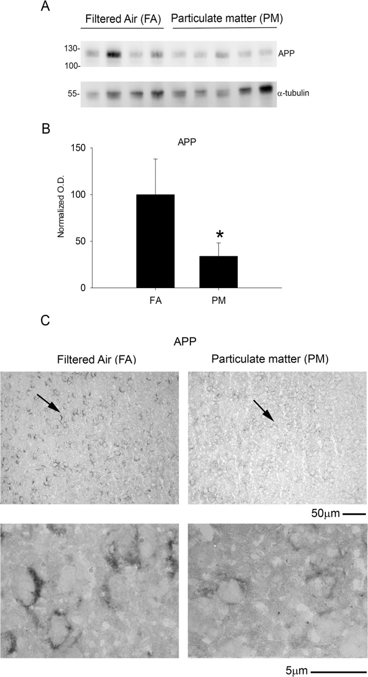
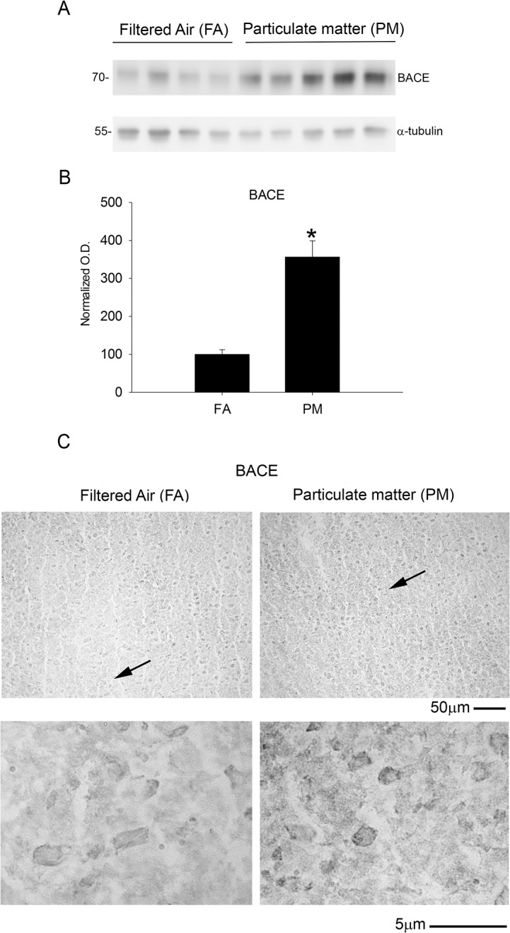
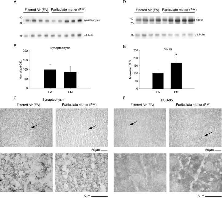
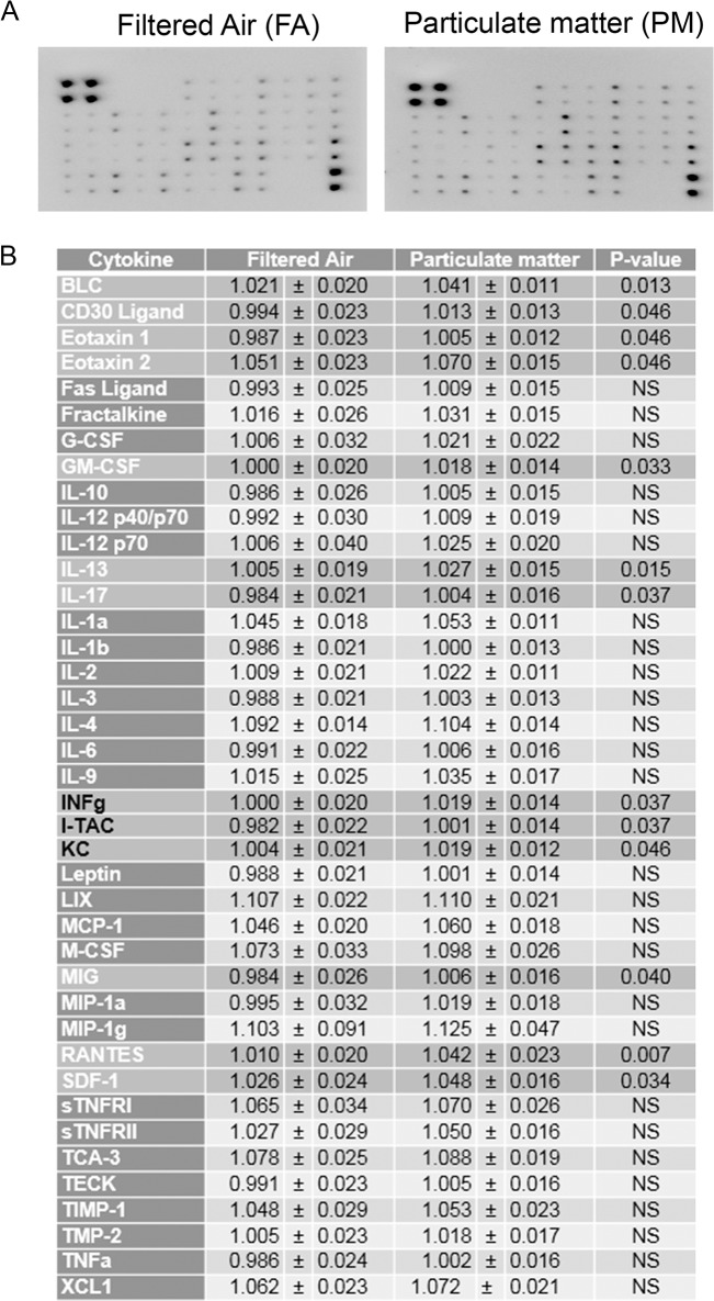
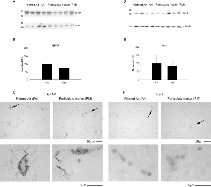
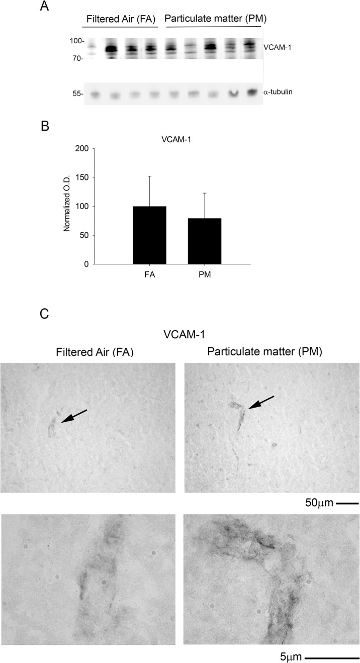
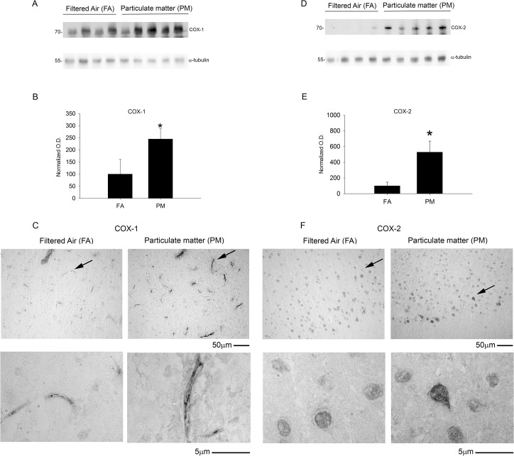
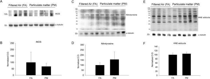
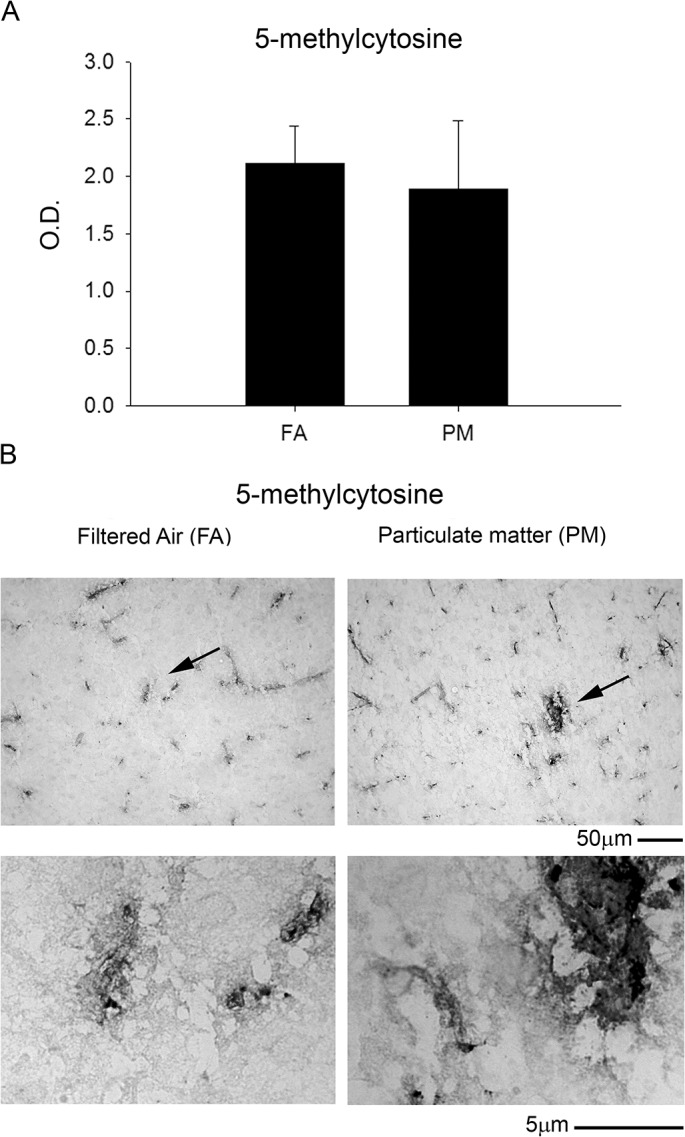
Exposure to air pollutants, including particulate matter, results in activation of the brain inflammatory response and Alzheimer disease (AD)-like pathology in dogs and humans. However, the length of time required for inhalation of ambient particulate matter to influence brain inflammation and AD pathology is less clear. Here, we studied the effect of 3 and 9 months of air particulate matter (<2.5 μm diameter, PM2.5) exposure on brain inflammatory phenotype and pathological hallmarks of AD in C57BL/6 mice. Using western blot, ELISA, and cytokine array analysis we quantified brain APP, beta-site APP cleaving enzyme (BACE), oligomeric protein, total Aβ 1-40 and Aβ 1-42 levels, inducible nitric oxide synthase (iNOS), nitrotyrosine-modified proteins, HNE-Michael adducts, vascular cell adhesion molecule 1 (VCAM-1), glial markers (GFAP, Iba-1), pre- and post- synaptic markers (synaptophysin and PSD-95), cyclooxygenase (COX-1, COX-2) levels, and the cytokine profile in PM2.5 exposed and filtered air control mice. Only 9 month PM2.5 exposure increased BACE protein levels, APP processing, and Aβ 1-40 levels. This correlated with a concomitant increase in COX-1 and COX-2 protein levels and a modest alteration in the cytokine profile. These data support the hypothesis that prolonged exposure to airborne particulate matter has the potential to alter brain inflammatory phenotype and promote development of early AD-like pathology.


Conflict of interest statement

Competing Interests: The authors have declared that no competing interests exist.

Figures

Fig 1
Fig 1. Brain amyloid beta 1–40 (Aβ 1–40) levels and Aβ immunoreactivity increased with 9 month airborne particulate matter exposure while no change in oligomerized protein staining was observed.
Brains of 9 month exposed filtered air (FA) or PM2.5 (PM) mice were collected with the left hemisphere fixed for immunohistochemistry and the right temporal cortices collected for biochemical analysis. A. Rodent Aβ 1–40 levels from FA and PM temporal cortices were quantified by ELISA, *p<0.05. B. Temporal cortex lysates from FA and PM brains were used for dot blot analysis using A11 anti-oligomer antibody and α-tubulin (loading control). C. A11 optical densities were normalized to their respective loading control, averaged (+/-SD), and graphed. Brains were immunostained using D, anti-A11 antibody to visualize oligomeric protein, E, anti-rodent specific Aβ antibody, and F, anti-Aβ antibody 4G8. Images shown are representative from the temporal cortex. Arrows indicate regions taken for higher magnification.
Fig 2
Fig 2. Brain microtubule-associated protein (tau) and its phosphorylated form (PHF-1) were not altered by 9 month exposure to airborne particulate matter.
Brains of 9 month exposed filtered air (FA) or PM2.5 (PM) mice were collected with the left hemisphere fixed for immunohistochemistry and the right temporal cortices collected for biochemical analysis. A. Temporal cortex lysates from FA and PM brains were used for western blot analysis using anti-phopsho tau (PHF-1), anti-tau (loading control for PHF-1), and anti-α-tubulin (loading control for tau), A11 anti-oligomer antibody and α-tubulin (loading control). B. PHF-1 optical densities were normalized to their tau loading controls, averaged (+/-SD), and graphed. C. Tau optical densities were normalized to their α-tubulin loading controls, averaged (+/-SD), and graphed.
Fig 3
Fig 3. Brain amyloid precursor protein (APP) levels were reduced after 9 month exposure to airborne particulate matter.
Brains of 9 month exposed filtered air (FA) or PM2.5 (PM) mice were collected with the left hemisphere fixed for immunohistochemistry and the right temporal cortices collected for biochemical analysis. A. Temporal cortex lysates from FA and PM brains were used for western blot analysis using anti-APP, Y188 antibody and α-tubulin (loading control). B. APP optical densities were normalized to their respective loading controls, averaged (+/-SD), and graphed, *p<0.05. C. Brains were immunostained using anti-APP, Y188 antibody. Images shown are representative from the temporal cortex. Arrows indicate regions taken for higher magnification.
Fig 4
Fig 4. Brain beta-site APP cleaving enzyme (BACE) protein levels increased after 9 month exposure to airborne particulate matter.
Brains of 9 month exposed filtered air (FA) or PM2.5 (PM) mice were collected with the left hemisphere fixed for immunohistochemistry and the right temporal cortices collected for biochemical analysis. A. Temporal cortex lysates from FA and PM brains were used for western blot analysis using anti-BACE antibody and α-tubulin (loading control). B. BACE optical densities were normalized to their respective loading controls, averaged (+/-SD), and graphed, *p<0.05. C. Brains were immunostained using anti-BACE antibody. Images shown are representative from the temporal cortex. Arrows indicate regions taken for higher magnification.
Fig 5
Fig 5. Brain post-synaptic marker, PSD-95, protein levels increased with 9 month airborne particulate matter exposure with no change in the pre-synaptic protein marker, synaptophysin.
Brains of 9 month exposed filtered air (FA) or PM2.5 (PM) mice were collected with the left hemisphere fixed for immunohistochemistry and the right temporal cortices collected for biochemical analysis. Temporal cortex lysates from FA and PM brains were used for western blot analysis using A, anti-synaptophysin, D, anti-PSD-95 and α-tubulin (loading control) antibodies. Optical densities for B, synaptophysin and E, PSD-95 were normalized to their respective loading controls, averaged (+/-SD), and graphed, *p<0.05. Brains were immunostained using C, anti-synaptophysin and F, anti-PSD-95 antibodies. Images shown are representative from the temporal cortex. Arrows indicate regions taken for higher magnification.
Fig 6
Fig 6. Brain inflammatory cytokines were modestly increased with 9 month airborne particulate matter exposure.
Brains of 9 month exposed filtered air (FA) or PM2.5 (PM) mice were collected with the left hemisphere fixed for immunohistochemistry and the right temporal cortices collected for biochemical analysis. A. Temporal cortex lysates from FA and PM brains were used for antibody-based commercial cytokine array analysis. A representative array is shown. B. The optical densities for each cytokine were averaged (+/-SD).
Fig 7
Fig 7. Brain astrocytic (GFAP) and microglial (Iba-1) protein markers were unaltered with 9 month airborne particulate matter exposure.
Brains of 9 month exposed filtered air (FA) or PM2.5 (PM) mice were collected with the left hemisphere fixed for immunohistochemistry and the right temporal cortices collected for biochemical analysis. Temporal cortex lysates from FA and PM brains were used for western blot analysis using A, anti-GFAP, D, anti-Iba-1 and α-tubulin (loading control) antibodies. Optical densities for B, GFAP and E, Iba-1 were normalized to their respective loading controls, averaged (+/-SD), and graphed. Brains were immunostained using C, anti-GFAP and F, anti-Iba-1 antibodies. Images shown are representative from the temporal cortex. Arrows indicate regions taken for higher magnification.
Fig 8
Fig 8. Vascular protein marker, VCAM-1, was unaltered with 9 month airborne particulate matter exposure.
Brains of 9 month exposed filtered air (FA) or PM2.5 (PM) mice were collected with the left hemisphere fixed for immunohistochemistry and the right temporal cortices collected for biochemical analysis. A. Temporal cortex lysates from FA and PM brains were used for western blot analysis using anti-VCAM antibody and α-tubulin (loading control). B. VCAM optical densities were normalized to their respective loading controls, averaged (+/-SD), and graphed. C. Brains were immunostained using anti-VCAM antibody. Images shown are representative from the temporal cortex. Arrows indicate regions taken for higher magnification.
Fig 9
Fig 9. Brain cyclooxygenase enzyme (COX-1 and COX-2) protein levels increased with 9 month airborne particulate matter exposure.
Brains of 9 month exposed filtered air (FA) or PM2.5 (PM) mice were collected with the left hemisphere fixed for immunohistochemistry and the right temporal cortices collected for biochemical analysis. Temporal cortex lysates from FA and PM brains were used for western blot analysis using A, anti-COX-1, D, anti-COX-2 and α-tubulin (loading control) antibodies. Optical densities for B, COX-1 and E, COX-2 were normalized to their respective loading controls, averaged (+/-SD), and graphed, *p<0.05. Brains were immunostained using C, anti-COX-1 and F, anti-COX-2 antibodies. Images shown are representative from the temporal cortex. Arrows indicate regions taken for higher magnification.
Fig 10
Fig 10. Brain oxidative and nitrosative stress protein marker levels were not altered with 9 month airborne particulate matter exposure.
Brains of 9 month exposed filtered air (FA) or PM2.5 (PM) mice were collected with the left hemisphere fixed for immunohistochemistry and the right temporal cortices collected for biochemical analysis. Temporal cortex lysates from FA and PM brains were used for western blot analysis using A, anti-inducible nitric oxide synthase (iNOS), C, nitrated tyrosines (nitrotyrosine), E, 4-hydroxynonenal protein Michael adducts (HNE adducts) and α-tubulin (loading control) antibodies. Optical densities for B, iNOS and E, nitrotyrosine, and F, HNE adducts were normalized to their respective loading controls, averaged (+/-SD), and graphed.
Fig 11
Fig 11. Brain DNA methylation was not altered with 9 month airborne particulate matter exposure.
Brains of 9 month exposed filtered air (FA) or PM2.5 (PM) mice were collected and the right parietal cortices were collected for DNA isolation. A. A 5-methylcytosine DNA ELISA was performed from isolated DNA (100ng/brain) from FA and PM brains. B. Optical densities were averaged (+/-SD) and graphed. C. Brains were immunostained using anti-5 methyl-cytosine antibody. Images shown are representative from the temporal cortex. Arrows indicate regions taken for higher magnification.

Similar articles

Cited by

References

    1. Maslow K, Maslow K, Alzheimer's A 2010 Alzheimer's disease facts and figures. Alzheimer's & dementia: the journal of the Alzheimer's Association. 2010;6: 158–94. - PubMed
    1. Thies W, Bleiler L, Alzheimer's A 2013 Alzheimer's disease facts and figures. Alzheimer's & dementia: the journal of the Alzheimer's Association. 2013;9: 208–45. - PubMed
    1. Pritchard C, Mayers A, Baldwin D Changing patterns of neurological mortality in the 10 major developed countries—1979-2010. Public health. 2013;127: 357–68. 10.1016/j.puhe.2012.12.018 - DOI - PubMed
    1. McGeer PL, McGeer EG The amyloid cascade-inflammatory hypothesis of Alzheimer disease: implications for therapy. Acta neuropathologica. 2013;126: 479–97. 10.1007/s00401-013-1177-7 - DOI - PubMed
    1. Cruchaga C, Kauwe JS, Harari O, Jin SC, Cai Y, Karch CM, et al. GWAS of cerebrospinal fluid tau levels identifies risk variants for Alzheimer's disease. Neuron. 2013;78: 256–68. 10.1016/j.neuron.2013.02.026 - DOI - PMC - PubMed

Publication types

MeSH terms