. 2015 May 19;42(5):815-25.
doi: 10.1016/j.immuni.2015.04.015.

Antibodies to Interleukin-2 Elicit Selective T Cell Subset Potentiation through Distinct Conformational Mechanisms

Affiliations

Antibodies to Interleukin-2 Elicit Selective T Cell Subset Potentiation through Distinct Conformational Mechanisms

Jamie B Spangler et al. Immunity. .

Abstract

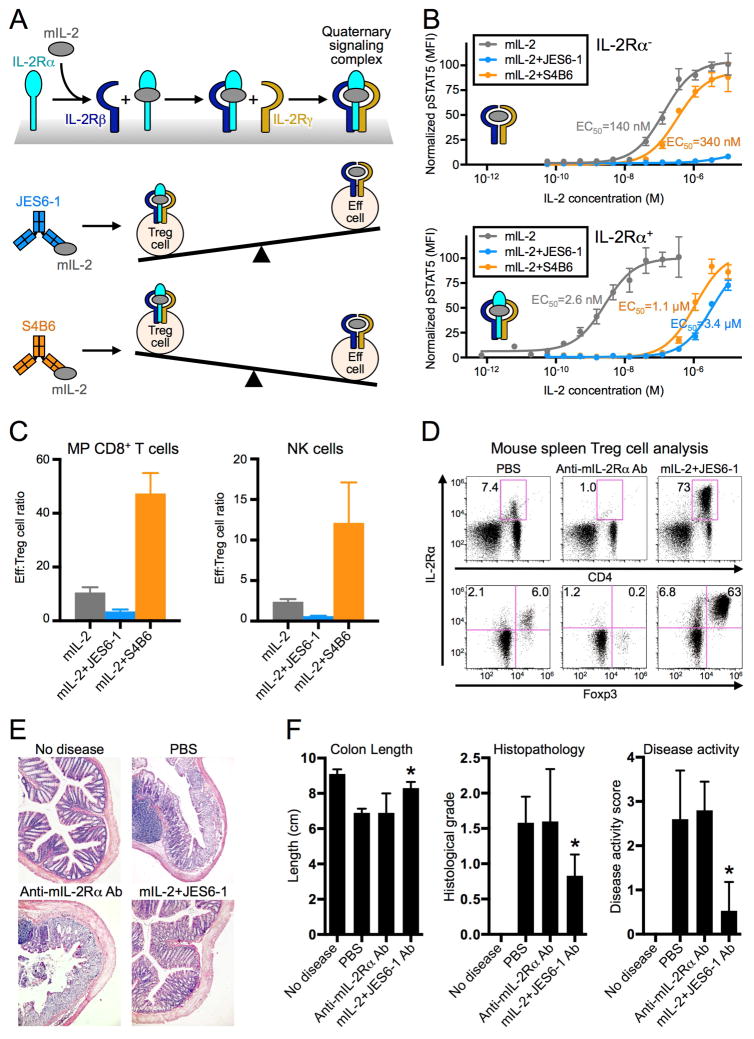
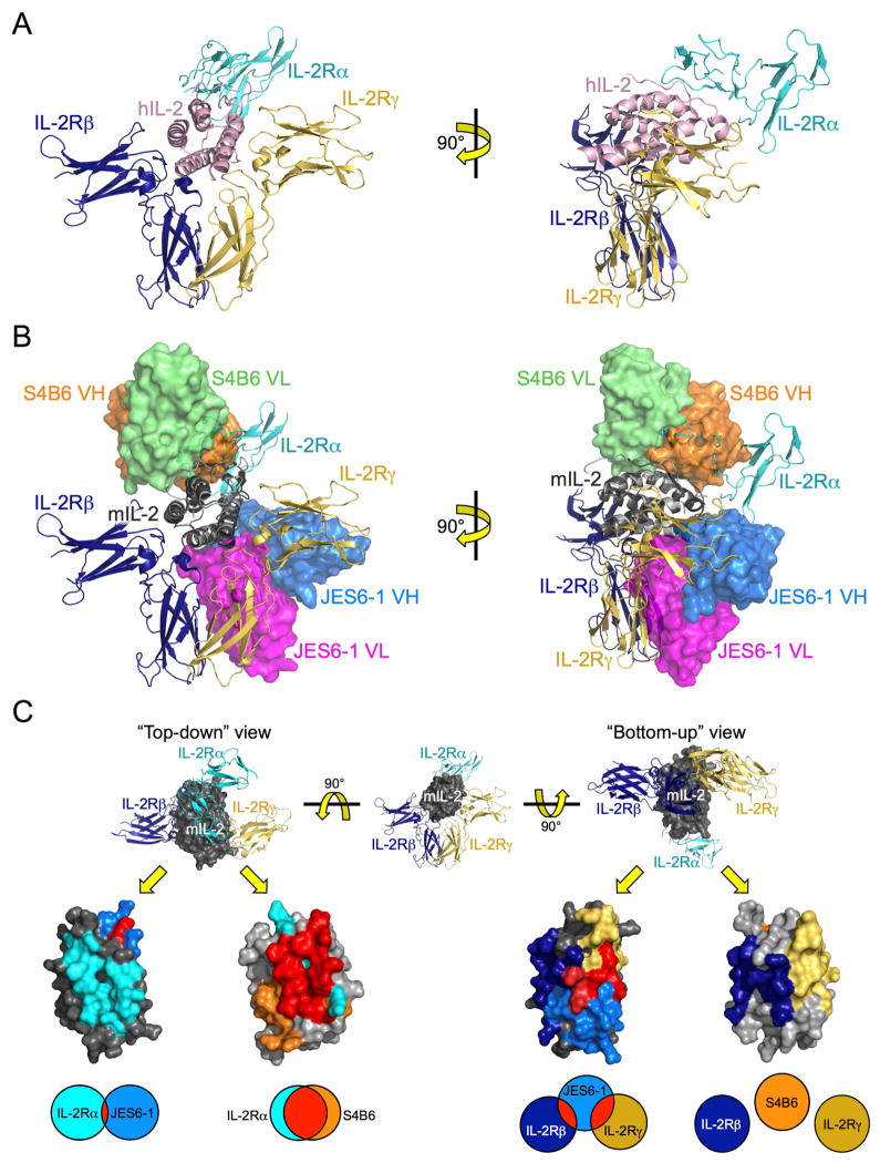
Interleukin-2 (IL-2) is a pleiotropic cytokine that regulates immune cell homeostasis and has been used to treat a range of disorders including cancer and autoimmune disease. IL-2 signals via interleukin-2 receptor-β (IL-2Rβ):IL-2Rγ heterodimers on cells expressing high (regulatory T cells, Treg) or low (effector cells) amounts of IL-2Rα (CD25). When complexed with IL-2, certain anti-cytokine antibodies preferentially stimulate expansion of Treg (JES6-1) or effector (S4B6) cells, offering a strategy for targeted disease therapy. We found that JES6-1 sterically blocked the IL-2:IL-2Rβ and IL-2:IL-2Rγ interactions, but also allosterically lowered the IL-2:IL-2Rα affinity through a "triggered exchange" mechanism favoring IL-2Rα(hi) Treg cells, creating a positive feedback loop for IL-2Rα(hi) cell activation. Conversely, S4B6 sterically blocked the IL-2:IL-2Rα interaction, while also conformationally stabilizing the IL-2:IL-2Rβ interaction, thus stimulating all IL-2-responsive immune cells, particularly IL-2Rβ(hi) effector cells. These insights provide a molecular blueprint for engineering selectively potentiating therapeutic antibodies.


Conflict of interest statement

The authors have no other competing financial interests.

Figures

Figure 1
Figure 1. Anti-IL-2 antibodies bias cytokine signaling and functional outcomes
(A) Schematic of IL-2 cytokine-receptor quaternary complex formation (top) and regulatory (Treg) versus effector (Eff) immune cell proliferation biases induced by mIL-2:JES6-1 (middle) and mIL-2:S4B6 (bottom) immunocomplexes. (B) STAT5 phosphorylation response to mIL-2 or mIL-2:antibody immunocomplex treatment in IL-2Ra (top) or IL-2Ra+ (bottom) YT-1 human NK cells. Half-maximal effective concentrations (EC50s) for STAT5 activation are indicated. Data are representative of three independent experiments. (C) C57BL/6 mouse spleen effector cell:Treg cell ratios following mIL-2 or immunocomplex treatment. Data are representative of two independent experiments. (D) Treg cell expansion in BALB/c mouse spleens in response to anti-mIL-2Rα antibody (Ab) or mIL-2:JES6-1 treatment. Top plots display the percentage of CD4+ cells that are IL-2Rα+. Bottom plots show only CD4+ cells and the percentages of Treg (IL-2Rα+FoxP3+) cells and IL-2Rα+FoxP3 cells are shown for each treatment condition. (E) Colon histopathology of colitis-induced BALB/c mice pretreated with anti-mIL-2Rα Ab or mIL-2:JES6-1. (F) Evaluation of disease progression indicators for treated mice. *P<0.05 by Student’s t-test (colon length) or Mann-Whitney U test (histological grade and disease activity score). All error bars indicate SD. Also see Figure S1.
Figure 2
Figure 2. mIL-2:JES6-1 and mIL-2:S4B6 complex structures reveal basis for competitive binding between anti-IL-2 antibodies and IL-2 receptor
(A) Orthosteric views of the hIL-2 cytokine-receptor quaternary complex (PDB ID 2B5I) comprised of hIL-2 (pink), IL-2Rα (cyan), IL-2Rβ (navy), and IL-2Rγ (gold). (B) Overlay of the mIL-2:JES6-1 and mIL-2:S4B6 complex structures on the hIL-2 quaternary complex. Both JES6-1-bound mIL-2 (dark gray) and S4B6-bound mIL-2 (light gray) are presented, with the JES6-1 variable heavy (VH, blue) and light (VL, magenta) and the S4B6 VH (orange) and VL (green) chains shown as surface representations. Crystallographic statistics for mIL-2:antibody complexes are provided in Table S1. (C) “Top-down” (left) and “bottom-up” (right) views of JES6-1-bound mIL-2 (dark gray) and S4B6-bound mIL-2 (light gray) with the predicted binding epitopes of IL-2Ra (cyan), IL-2Rβ (navy), IL-2R (gold), JES6-1 (blue), and S4B6 (orange) on the mIL-2 cytokine shaded. Residues shared between the mIL-2:antibody and the mIL-2:IL-2Ra, mIL-2:IL-2Rβ, or mIL-2:IL-2R interfaces are colored red. Venn diagrams at bottom indicate the relative extent of overlap between the antibody and receptor epitopes. Also see Figure S2.
Figure 3
Figure 3. JES6-1 allosterically obstructs IL-2Rα binding to IL-2 via epitope distortion
Overlay of the hIL-2 quaternary complex (PDB ID 2B5I) with the mIL-2:JES6-1 complex. Surface depictions of the IL-2Rα (cyan), IL-2Rβ (navy), and IL-2Rγ (gold) subunits and the JES6-1 antibody (blue) and cartoon representations of receptor-bound hIL-2 (pink) and JES6-1-bound mIL-2 (red) are shown. An enlargement of the IL-2Rα binding site is provided at right with key IL-2Ra-interacting residues in the hIL-2 AB loop and the corresponding JES6-1-bound mIL-2 residues labeled. Arrows highlight differences between the structures. Also see Figure S3.
Figure 4
Figure 4. S4B6 mimics the IL-2Ra subunit, allosterically enhancing binding of IL-2Rβ
(A) Overlay of hIL-2 (pink) bound to IL-2Rα (cyan), IL-2Rβ (navy), and IL-2Rγ and mIL-2 (light gray) bound to S4B6 (orange) with detailed views of the hIL-2:hIL-2Ra (left) and mIL-2:S4B6 (right) interfaces in the AB loops and B helices of the cytokines juxtaposed at right. Analogous human and mouse IL-2 residues implicated in both interactions are labeled. (B) Superposition of cytokine orientations proximal to the IL-2Rβ binding site for unbound super-2 (PDB ID 3QB1, salmon), receptor-bound hIL-2 from the quaternary complex (PDB ID 2B5I, pink), S4B6-bound mIL-2 (red), and unbound hIL-2 (PDB ID 3INK, green). Also see Figure S4.
Figure 5
Figure 5. Anti-IL-2 antibodies and IL-2 receptor compete for cytokine binding
Yeast surface mIL-2 competition studies between anti-mIL-2 antibodies (Abs) and saturating concentrations of mIL-2Rα (A) or mIL-2Rβ (B) are shown (Error bars, SD). Data are representative of three independent experiments. Half maximal inhibitory concentrations (IC50s) of the antibodies are indicated. Equilibrium surface plasmon resonance titrations of mIL-2:antibody immunocomplex interactions with immobilized mIL-2Rα (C) or mIL-2Rβ (D) and mIL-2Rα (E) or mIL-2Rβ (F) interactions with unbound mIL-2 compared to interactions with immobilized mIL-2:antibody immunocomplexes are presented. Also see Figure S5.
Figure 6
Figure 6. mIL-2:JES6-1 immunocomplex induces increased IL-2Rα expression to create a positive feedback loop favoring IL-2Rαhi cell signaling
(A) IL-2Rα expression quantification (mean fluorescence intensity, MFI) for five immune cell subsets (Treg, MP CD8+ T, NK, NKT, and γd TCR) in BALB/c mouse spleens following treatment with mIL-2 or mIL-2:antibody immunocomplex. (B) Percentage of IL-2Rα+ cells within each immune cell subset in treated mouse spleens. (C) IL-2Rα expression histograms for Treg cells isolated from treated mouse spleens. Representative plots from one mouse per cohort are presented. (D) Relative cell expansion (transparent thick bars) and IL-2Rα expression quantification (solid thin bars) in treated mouse spleens. Responses for each treatment cohort are normalized to those of untreated control mice for each cell subset. (E) Relative cell expansion (transparent thick bars) and IL-2Rα+ cell percentage (solid thin bars) in treated mouse spleens, normalized as in (D). Error bars indicate SD. Data are representative of two independent experiments.

Comment in

References

    1. Adams PD, Grosse-Kunstleve RW, Hung LW, Ioerger TR, McCoy AJ, Moriarty NW, Read RJ, Sacchettini JC, Sauter NK, Terwilliger TC. PHENIX: building new software for automated crystallographic structure determination. Acta Crystallogr D Biol Crystallogr. 2002;58:1948–1954. - PubMed
    1. Boder ET, Wittrup KD. Yeast surface display for screening combinatorial polypeptide libraries. Nature biotechnology. 1997;15:553–557. - PubMed
    1. Boyman O, Kovar M, Rubinstein MP, Surh CD, Sprent J. Selective stimulation of T cell subsets with antibody-cytokine immune complexes. Science. 2006a;311:1924–1927. - PubMed
    1. Boyman O, Sprent J. The role of interleukin-2 during homeostasis and activation of the immune system. Nat Rev Immunol. 2012;12:180–190. - PubMed
    1. Boyman O, Surh CD, Sprent J. Potential use of IL-2/anti-IL-2 antibody immune complexes for the treatment of cancer and autoimmune disease. Expert Opin Biol Ther. 2006b;6:1323–1331. - PubMed

Publication types

MeSH terms