

Image-Guided Radiotherapy Using a Modified Industrial Micro-CT for Preclinical Applications
- PMID: 25993010
- PMCID: PMC4438006
- DOI: 10.1371/journal.pone.0126246
Image-Guided Radiotherapy Using a Modified Industrial Micro-CT for Preclinical Applications
Abstract
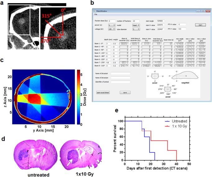
Purpose/objective: Although radiotherapy is a key component of cancer treatment, its implementation into pre-clinical in vivo models with relatively small target volumes is frequently omitted either due to technical complexity or expected side effects hampering long-term observational studies. We here demonstrate how an affordable industrial micro-CT can be converted into a small animal IGRT device at very low costs. We also demonstrate the proof of principle for the case of partial brain irradiation of mice carrying orthotopic glioblastoma implants.
Methods/materials: A commercially available micro-CT originally designed for non-destructive material analysis was used. It consists of a CNC manipulator, a transmission X-ray tube (10-160 kV) and a flat-panel detector, which was used together with custom-made steel collimators (1-5 mm aperture size). For radiation field characterization, an ionization chamber, water-equivalent slab phantoms and radiochromic films were used. A treatment planning tool was implemented using a C++ application. For proof of principle, NOD/SCID/γc(-/-) mice were orthotopically implanted with U87MG high-grade glioma cells and irradiated using the novel setup.
Results: The overall symmetry of the radiation field at 150 kV was 1.04 ± 0.02%. The flatness was 4.99 ± 0.63% and the penumbra widths were between 0.14 mm and 0.51 mm. The full width at half maximum (FWHM) ranged from 1.97 to 9.99 mm depending on the collimator aperture size. The dose depth curve along the central axis followed a typical shape of keV photons. Dose rates measured were 10.7 mGy/s in 1 mm and 7.6 mGy/s in 5 mm depth (5 mm collimator aperture size). Treatment of mice with a single dose of 10 Gy was tolerated well and resulted in central tumor necrosis consistent with therapeutic efficacy.
Conclusion: A conventional industrial micro-CT can be easily modified to allow effective small animal IGRT even of critical target volumes such as the brain.
Conflict of interest statement
Figures

References
-
- Graves EE, Zhou H, Chatterjee R, Keall PJ, Gambhir SS, Contag CH, et al. Design and evaluation of a variable aperture collimator for conformal radiotherapy of small animals using a microCT scanner. Med Phys 2007;34:4359–4367. - PubMed
Publication types
MeSH terms
LinkOut - more resources
Full Text Sources
Other Literature Sources
