. 2015 May 20:5:10223.
doi: 10.1038/srep10223.

Magnetic resonance enterographic predictors of one-year outcome in ileal and ileocolonic Crohn's disease treated with anti-tumor necrosis factor antibodies

Affiliations

Magnetic resonance enterographic predictors of one-year outcome in ileal and ileocolonic Crohn's disease treated with anti-tumor necrosis factor antibodies

Piotr Eder et al. Sci Rep. .

Abstract

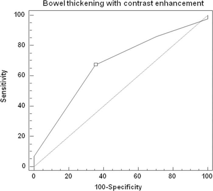
The aim of the study was to assess the role of magnetic resonance enterography (MRE) in predicting one-year efficacy of anti-tumor necrosis factor antibodies--infliximab (IFX), adalimumab (ADA) in Crohn's disease (CD) patients primarily responding to therapy. We performed retrospective analysis among 61 CD patients who had undergone a successful IFX/ADA induction therapy and were treated with maintenance doses. All patients underwent MRE at week 0. We assessed which MRE features were predictive for steroid-free remission at week 52, and which were associated with a secondary loss of response. 44 patients were in steroid-free remission at week 52, 17--were secondary non-responders. The ROC curve showed that bowel thickening with contrast enhancement analyzed together at week 0 were associated with steroid-free remission at week 52 (p = 0.01; AUC 0.67). Bowel stenosis with or without prestenotic dilatation [OR 5.8 (95% CI 1.4-25) and 2.4 (95% CI 1.2 - 5) respectively; p = 0.01] and the presence of intra-abdominal fistulas [OR 1.4 (95% CI 1.1-2); p=0.004] were related to secondary non-response. A high baseline inflammatory activity detected by MRE predicts one-year response in CD after IFX/ADA. In case of bowel stenosis, intra-abdominal fistulas, other therapeutic options should be considered.


Conflict of interest statement

PE, LLS, and KK received lecture fees from Abbvie Poland. Other authors declare no competing financial interests.

Figures

Figure 1
Figure 1
Receiver operator characteristic (ROC) curve showing that the score assessing bowel inflammatory thickening with contrast enhancement in the baseline magnetic resonance enterography (range 0 – 4 points) was predictive for long-term steroid-free remission at week 52 in Crohn’s disease patients treated with anti-TNF antibodies. An optimal cut-off point was ≥ 3, and it allowed to predict with 67% (confidence interval: 52 – 81%) sensitivity and 65% (confidence interval: 39 – 86%) specificity achievement of the primary therapeutic end point of the study (p = 0.01; area under the curve 0.67).
Figure 2
Figure 2
Magnetic resonance enterography images illustrating an example of a response to one-year anti-tumor necrosis factor (anti-TNF) therapy in a patient with Crohn’s disease: (a) Week 0 - before anti-TNF therapy. T2-weighted sequence demonstrating thickening of a bowel wall (white arrows) without stenosis of the bowel lumen. (b) Week 0 - before anti-TNF therapy. Dynamic contrast enhanced T1-volume interpolated gradient-echo sequence demonstrating thickening of a bowel wall with enhancing in the various layers of the wall - hyperintense signal corresponding to edema in acute inflammation (white arrows). (c) Week 52. T2-weighted sequence demonstrating decrease of thickening of the bowel wall (white arrows) after one-year anti-TNF therapy. (d) Week 52. Dynamic contrast enhanced T1-volume interpolated gradient-echo sequence demonstrating decrease of thickening of the bowel wall with moderate enhancement typical for moderate activity of inflammation (white arrows) - after one-year anti-TNF therapy.
Figure 3
Figure 3
Magnetic resonance enterography images illustrating an example of a non-response to one-year anti-tumor necrosis factor (anti-TNF) therapy in a patient with Crohn’s disease: (a) Week 0 - before anti-TNF therapy. T2-weighted sequence demonstrating thickening of a bowel wall (white arrows) with stenosis of the bowel lumen with prestenotic dilatation (asterisk). (b) Week 0 - before anti-TNF therapy. Dynamic contrast enhanced T1-volume interpolated gradient-echo sequence demonstrating thickening of a bowel wall with enhancing in the various layers of the wall - hyperintense signal corresponding to edema in acute inflammation and dilatation of lumen (white arrows). (c) Week 52. T2-weighted sequence demonstrating increase of thickening of the bowel wall (white arrow) with stenosis of lumen and prestenotic dilatation (asterisk) after one-year anti-TNF therapy. (d) Week 52. Dynamic contrast enhanced T1-volume interpolated gradient-echo sequence demonstrating increase of thickening of the bowel wall with enhancement typical for activity of inflammation (white arrows) – after one-year anti-TNF therapy.

References

    1. Feagan B. G. et al. Recommendations for the treatment of Crohn’s disease with tumor necrosis factor antagonists: an expert consensus report. Inflamm. Bowel Dis. 1, 152–160 (2012). - PubMed
    1. Allez M. et al. Report of the ECCO pathogenesis workshop on anti-TNF therapy failures in inflammatory bowel diseases: definitions, frequency and pharmacological aspects. J. Crohns Colitis 4, 355–366 (2010). - PubMed
    1. Vermeire S. & Gils A. Value of drug level testing and antibody assays in optimizing biological therapy. Frontline Gastroenterology 4, 41–43 (2013). - PMC - PubMed
    1. Siegel C. A. & Melmed G. Y. Predicting response to anti-TNF agents for the treatment of Crohn’s disease. Ther. Adv. Gastroenterol. 2, 245–251 (2009). - PMC - PubMed
    1. Amitai M., Ben-Horin S., Eliakim R. & Kopylow U. Magnetic resonance enterography in Crohn’s disease: a guide to common imaging manifestations for the IBD physician. J. Crohns Colitis 8, 603–615 (2013). - PubMed