. 2015 May 20;10(5):e0127691.
doi: 10.1371/journal.pone.0127691. eCollection 2015.

Novel Features of the Prenatal Horn Bud Development in Cattle (Bos taurus)

Affiliations

Novel Features of the Prenatal Horn Bud Development in Cattle (Bos taurus)

Dominique Judith Wiener et al. PLoS One. .

Abstract

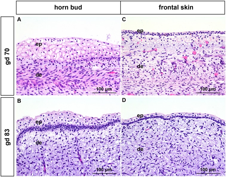
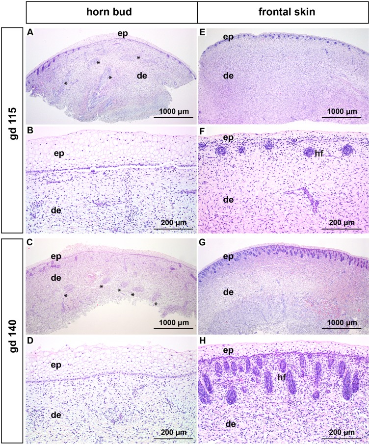
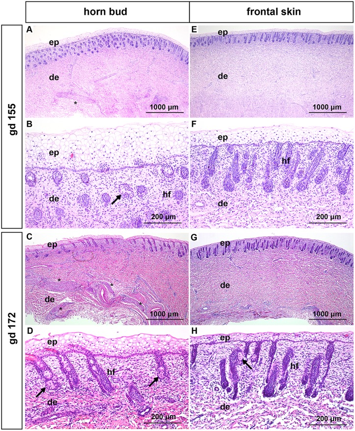
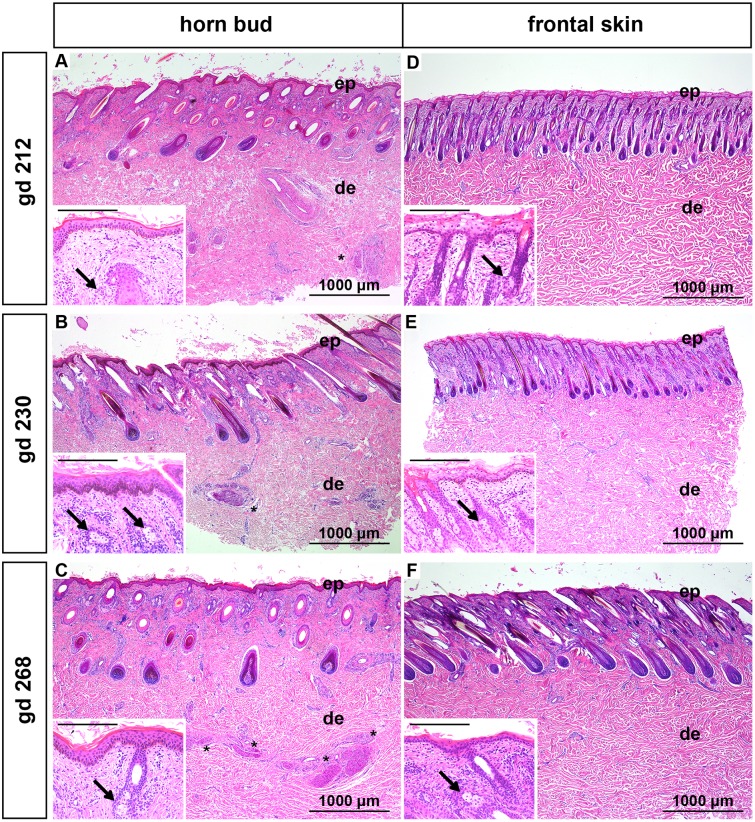
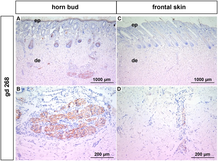
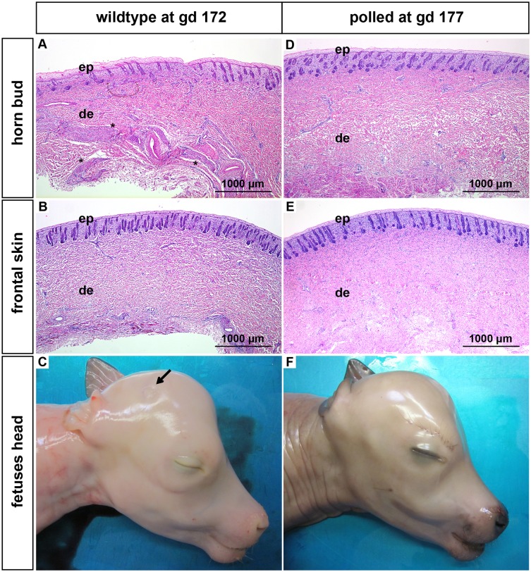
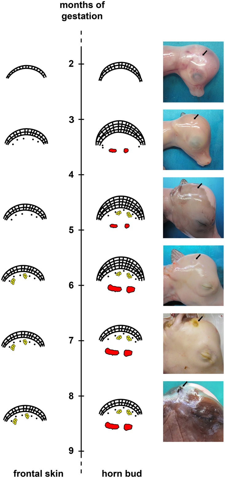
Whereas the genetic background of horn growth in cattle has been studied extensively, little is known about the morphological changes in the developing fetal horn bud. In this study we histologically analyzed the development of horn buds of bovine fetuses between ~70 and ~268 days of pregnancy and compared them with biopsies taken from the frontal skin of the same fetuses. In addition we compared the samples from the wild type (horned) fetuses with samples taken from the horn bud region of age-matched genetically hornless (polled) fetuses. In summary, the horn bud with multiple layers of vacuolated keratinocytes is histologically visible early in fetal life already at around day 70 of gestation and can be easily differentiated from the much thinner epidermis of the frontal skin. However, at the gestation day (gd) 212 the epidermis above the horn bud shows a similar morphology to the epidermis of the frontal skin and the outstanding layers of vacuolated keratinocytes have disappeared. Immature hair follicles are seen in the frontal skin at gd 115 whereas hair follicles below the horn bud are not present until gd 155. Interestingly, thick nerve bundles appear in the dermis below the horn bud at gd 115. These nerve fibers grow in size over time and are prominent shortly before birth. Prominent nerve bundles are not present in the frontal skin of wild type or in polled fetuses at any time, indicating that the horn bud is a very sensitive area. The samples from the horn bud region from polled fetuses are histologically equivalent to samples taken from the frontal skin in horned species. This is the first study that presents unique histological data on bovine prenatal horn bud differentiation at different developmental stages which creates knowledge for a better understanding of recent molecular findings.


Conflict of interest statement

Competing Interests: The authors have declared that no competing interests exist.

Figures

Fig 1
Fig 1. Histological analysis of horn buds and frontal skin from wild type fetuses (gd 70 and 83).
(A) and (B): Horn buds with multiple layers of vacuolated keratinocytes. (C) and (D): Frontal skin. Haematoxylin and eosin. Gd = gestation days, ep = epidermis, de = dermis.
Fig 2
Fig 2. Histological analysis of horn buds and frontal skin from wild type fetuses (gd 115 and 140).
(A-D): Horn buds with multiple layers of vacuolated keratinocytes. Note the absence of hair follicles below the horn bud in (A-D). (A) and (C): Note thick nerve bundles in the dermis below the horn bud (black stars). (B) and (D): Represent magnifications (A) and (C). (E-H): Frontal skin. (E) and (G): Note the absence of thick nerve bundles in the dermis. (F) and (H) represent magnifications of (E) and (G). Haematoxylin and eosin. Gd = gestation days, ep = epidermis, de = dermis, hf = hair follicles.
Fig 3
Fig 3. Histological analysis of horn buds and frontal skin from wild type fetuses (gd 155 and172).
(A-D): Horn buds with multiple layers of vacuolated keratinocytes. (A) and (C): Note thick nerve bundles in the dermis below the horn bud (black stars). (B) and (D): Represent magnifications of (A) and (C). Note presence of sebaceous glands (black arrows). (E-H): Frontal skin. (E) and (G): Note the absence of thick nerve bundles in the dermis. (F) and (H): Represent magnifications of (E) and (G). (H): Note the presence of sebaceous glands (black arrows). Haematoxylin and eosin. Gd = gestation days, ep = epidermis, de = dermis, hf = hair follicles.
Fig 4
Fig 4. Histological analysis of horn buds and frontal skin from wild type fetuses (gd 212, 230 and 268).
(A-C): Horn buds. Note differentiated epidermis and thick nerve bundles in the dermis below the horn bud (black stars). Insets show well-differentiated keratinocytes in the epidermis and sebaceous glands (black arrows). (D-F): Frontal skin. Note the absence of thick nerve bundles in the dermis. Insets show immature keratinocytes in (D) and (E) and well-differentiated keratinocytes in (F). Note sebaceous glands in the insets (black arrows). Haematoxylin and eosin. Insets: Scale bar represents 200μm. Gd = gestation days, ep = epidermis, de = dermis.
Fig 5
Fig 5. Neuron specific enolase staining from a wild type fetus (gd 268).
(A) and (B): Horn bud. (B): Magnification of (A). Note positive staining of the thick nerve bundles in the dermis. (C) and (D): Frontal skin. (D): Magnification of (C). Note positive staining of nerve fibres of normal size in the dermis. Gd = gestation days, ep = epidermis, de = dermis.
Fig 6
Fig 6. Features of horn buds and frontal skin from wild type and polled fetuses (gd 172 and 177).
(A): Horn bud from a wild type fetus with multiple layers of vacuolated keratinocytes. Note presence of thick nerve bundles in the dermis below the horn bud (black stars). (B): Frontal skin from a wild type fetus. Note absence of thick nerve bundles in the dermis. (C): Macroscopic picture of a horn bud from a wild type fetus. Note indentation of skin (black arrow). Region of the horn bud (D) and frontal skin (E) from a polled fetus. Note absence of thick nerve bundles in the dermis. (F): Macroscopic picture of a polled fetus without indentation of the skin. Haematoxylin and eosin. Gd = gestation days, ep = epidermis, de = dermis.
Fig 7
Fig 7. Schematic illustration of developmental stages of the horn bud and frontal skin in wild type fetuses.
Horn bud: Multiple layers of keratinocytes are present between 2 and 6 months of gestation and hair follicles are lacking below the horn bud between 2 and 4 months. Note the appearance of thick nerve bundles below the horn bud at 3 months of gestation. Hair follicles and sebaceous glands are present below the horn bud at 4–5 months. Frontal skin: Note thin epidermis at all time-points. Sebaceous glands are present at about 5–6 months of gestation. Note absence of thick nerve bundles in the dermis. Macroscopic pictures: The black arrow depicts the region of the horn bud. Note presence of hair follicles at 6–7 months of gestation. Red = nerve fibres, yellow = sebaceous glands.

References

    1. Dyce KM, Sack WC, Wensing CJG. The common integument In: Dyce KM, Sack WC, Wensing CJG, editors. Textbook of veterinary anatomy; 2002. p. 359.
    1. Reese S, Budras KD, Muelling C, Bragulla H, Koenig HE. Allgemeine Körperdecke (Integumentum Commune) In: König HE, Liebich HG, editors. Anatomie der Haussäugetiere. Schattauer; 2011. 653–655 pp.
    1. Strouhal E. Life of the Ancient Egyptians. 1st ed Liverpool University Press; 1997. 128 p.
    1. Graf B, Senn M. Behavioural and physiological responses of calves to dehorning by heat cauterization with or without local anaesthesia. Appl Anim Behav Sci. 1999; 62: 153–171.
    1. Alsaaod M, Doherr MG, Greber D, Steiner A. Experience with the delegation of anaesthesia for disbudding and castration to trained and certified livestock owners. BMC Vet Res. 2014; 10: 35 10.1186/1746-6148-10-35 - DOI - PMC - PubMed

Publication types

Substances