. 2015 Dec;25(12):3480-7.
doi: 10.1007/s00330-015-3764-6. Epub 2015 May 21.

Prevalence of cartilaginous tumours as an incidental finding on MRI of the knee

Collaborators, Affiliations

Prevalence of cartilaginous tumours as an incidental finding on MRI of the knee

Wouter Stomp et al. Eur Radiol. 2015 Dec.

Abstract

Objectives: The purpose was to determine prevalence of enchondromas and atypical cartilaginous tumour/chondrosarcoma grade 1 (ACT/CS1) of the knee on MRI in a large cohort study, namely the Netherlands Epidemiology of Obesity (NEO) study.

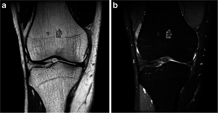
Methods: Participants aged 45 to 65 years were prospectively included, oversampling overweight and obese persons. Within a subgroup of participants, MRI of the right knee was performed and screened for incidental cartilaginous tumours, as defined by their characteristic location and appearance.

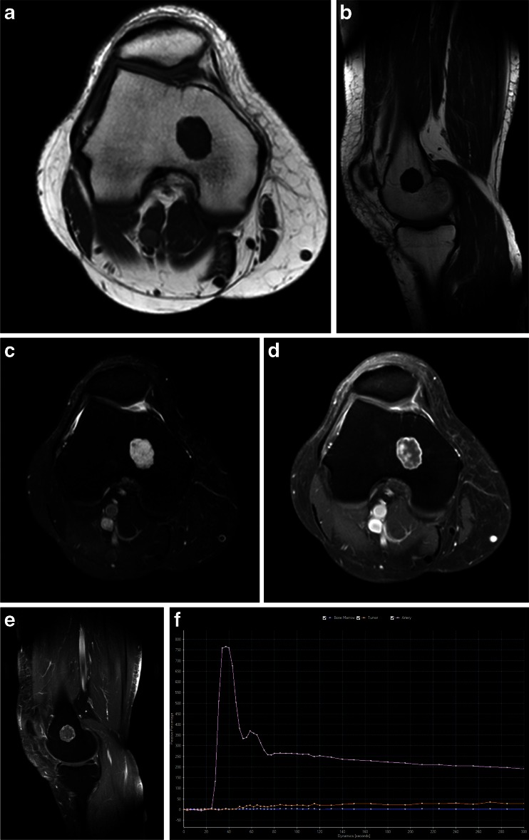
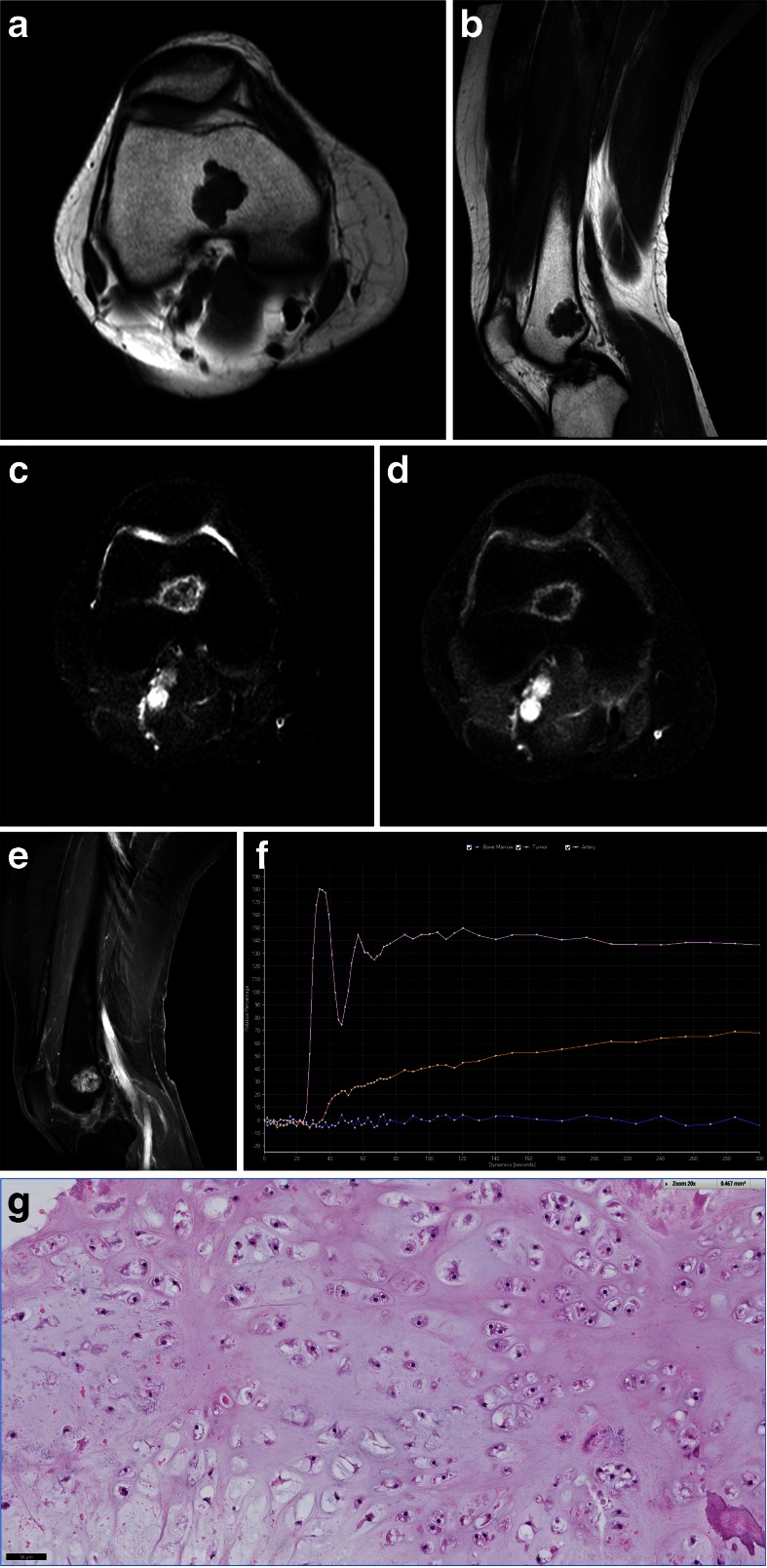
Results: Forty-nine cartilaginous tumours were observed in 44 out of 1285 participants (estimated population prevalence 2.8 %, 95 % CI 2.0-4.0 %). Mean largest tumour diameter was 12 mm (range 2-31 mm). Eight participants with a tumour larger than 20 mm or a tumour with aggressive features were referred to rule out low-grade chondrosarcoma. One was lost to follow-up, three had histologically proven ACT/CS1 and four had dynamic contrast MRI findings consistent with benign enchondroma.

Conclusions: Incidental cartilaginous tumours were relatively common on knee MRI and may be regarded as a normal concurrent finding. However, more tumours than expected were ACT/CS1. Because further examination was performed only when suspicion of chondrosarcoma was high, the actual prevalence might be even higher.

Key points: • Incidental cartilaginous tumours are relatively common on knee MRI. • Most incidental cartilaginous tumours are small and lack suspicious features. • Small cartilaginous tumours without suspicious findings may be a normal concurrent finding. • Large tumours and/or those with suspicious findings should be further investigated. • Atypical cartilaginous tumour/chondrosarcoma grade 1 was found more often than expected.

Keywords: Chondroma; Chondrosarcoma; Incidental findings; Knee; Magnetic resonance imaging.


Figures

Fig. 1
Fig. 1
Small enchondroma in the distal femur located centrally in the metaphysis and with a typical lobulated appearance: a PD-weighted coronal section, b PD-weighted coronal section with fat saturation. No further imaging was obtained
Fig. 2
Fig. 2
Large cartilaginous tumour centrally located in the distal femur, referred for follow-up: a axial and b sagittal T1-weighted images, c axial T2-weighted image, d axial and e sagittal T1-weighted images after gadolinium administration with fat suppression. The time–signal intensity curve of the dynamic MRI (e) showed slow enhancement consistent with enchondroma (pink artery, orange tumour, and blue bone marrow as reference tissue). The vertical axis represents relative signal intensity and the horizontal axis represents time in seconds
Fig. 3
Fig. 3
Large cartilaginous tumour centrally located in the distal femur, referred for follow-up: a axial and b sagittal T1-weighted images, c axial T2-weighted image, d axial and e sagittal T1-weighted images after gadolinium administration with fat suppression. The time–signal intensity curve of the dynamic MRI (e) showed fast enhancement (<10 s), interpreted as most likely ACT/CS1, although chondrosarcoma grade 2 could not be excluded due to extensive presence of mucoid (pink artery, orange tumour, and blue bone marrow as reference tissue). The vertical axis represents relative signal intensity and the horizontal axis represents time in seconds. Curettage was performed and histology (f) confirmed the presence of cartilaginous tumour with increased cellularity and occasional binucleated cells as well as focal mucomyxoid matrix changes (left lower area), diagnosed as atypical cartilaginous tumour/chondrosarcoma grade 1

References

    1. Kransdorf MJ, Peterson JJ, Bancroft LW. MR imaging of the knee: incidental osseous lesions. Magn Reson Imaging Clin N Am. 2007;15:13–24. doi: 10.1016/j.mric.2007.02.002. - DOI - PubMed
    1. Flemming DJ, Murphey MD. Enchondroma and chondrosarcoma. Semin Musculoskelet Radiol. 2000;4:59–71. doi: 10.1055/s-2000-6855. - DOI - PubMed
    1. Milgram JW. The origins of osteochondromas and enchondromas. A histopathologic study. Clin Orthop Relat Res. 1983;174:264–284. - PubMed
    1. Amary MF, Bacsi K, Maggiani F, et al. IDH1 and IDH2 mutations are frequent events in central chondrosarcoma and central and periosteal chondromas but not in other mesenchymal tumours. J Pathol. 2011;224:334–343. doi: 10.1002/path.2913. - DOI - PubMed
    1. Pansuriya TC, van Eijk R, d’ Adamo P, et al. Somatic mosaic IDH1 and IDH2 mutations are associated with enchondroma and spindle cell hemangioma in Ollier disease and Maffucci syndrome. Nat Genet. 2011;43:1256–1261. doi: 10.1038/ng.1004. - DOI - PMC - PubMed

Publication types