

Modulation of cell function by electric field: a high-resolution analysis
- PMID: 25994294
- PMCID: PMC4590499
- DOI: 10.1098/rsif.2015.0153
Modulation of cell function by electric field: a high-resolution analysis
Abstract
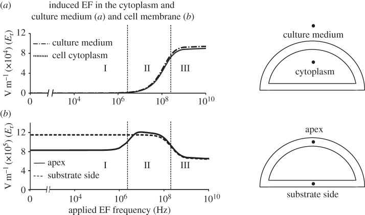
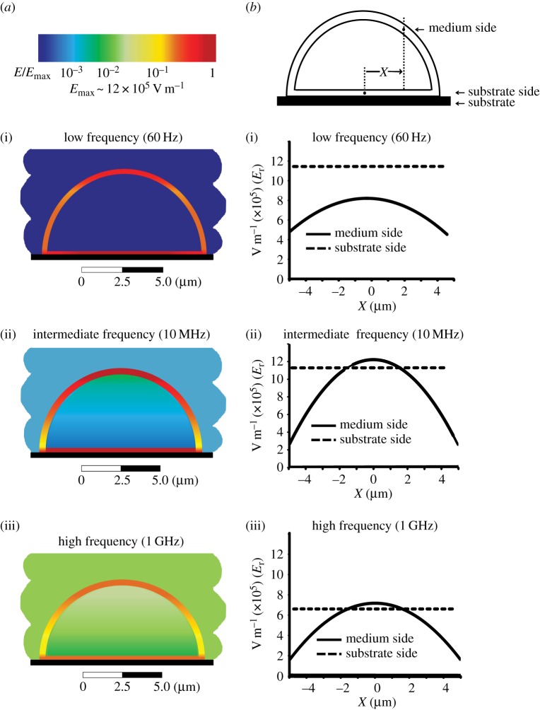
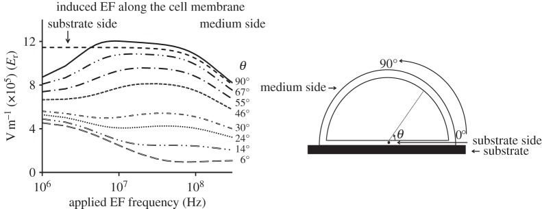
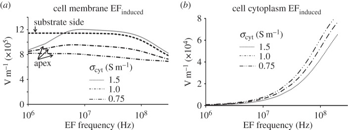
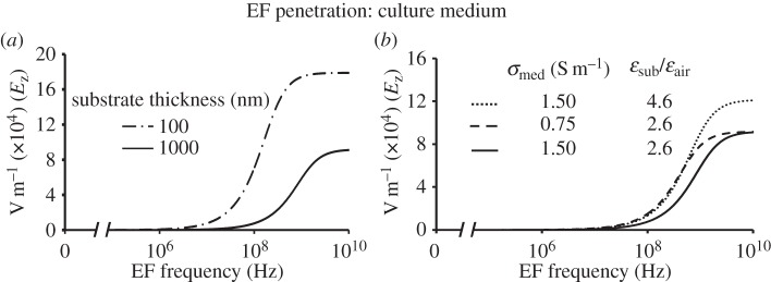
Regulation of cell function by a non-thermal, physiological-level electromagnetic field has potential for vascular tissue healing therapies and advancing hybrid bioelectronic technology. We have recently demonstrated that a physiological electric field (EF) applied wirelessly can regulate intracellular signalling and cell function in a frequency-dependent manner. However, the mechanism for such regulation is not well understood. Here, we present a systematic numerical study of a cell-field interaction following cell exposure to the external EF. We use a realistic experimental environment that also recapitulates the absence of a direct electric contact between the field-sourcing electrodes and the cells or the culture medium. We identify characteristic regimes and present their classification with respect to frequency, location, and the electrical properties of the model components. The results show a striking difference in the frequency dependence of EF penetration and cell response between cells suspended in an electrolyte and cells attached to a substrate. The EF structure in the cell is strongly inhomogeneous and is sensitive to the physical properties of the cell and its environment. These findings provide insight into the mechanisms for frequency-dependent cell responses to EF that regulate cell function, which may have important implications for EF-based therapies and biotechnology development.
Keywords: cell transmembrane potential; cell-substrate interaction; electrical cell stimulation; frequency-dependent response; surface charge.
© 2015 The Author(s) Published by the Royal Society. All rights reserved.
Figures
Similar articles
-
Biological cell response to electric field: a review of equivalent circuit models and future challenges.Biomed Phys Eng Express. 2025 Jan 24;11(2). doi: 10.1088/2057-1976/ad8092. Biomed Phys Eng Express. 2025. PMID: 39332436 Review.
-
Effect of External Electric Field on Substrate Transport of a Secondary Active Transporter.J Chem Inf Model. 2016 Aug 22;56(8):1539-46. doi: 10.1021/acs.jcim.6b00212. Epub 2016 Jul 29. J Chem Inf Model. 2016. PMID: 27472561
-
On-chip dielectrophoretic coassembly of live cells and particles into responsive biomaterials.Langmuir. 2010 Mar 2;26(5):3441-52. doi: 10.1021/la902989r. Langmuir. 2010. PMID: 19957941
-
Electrophoresis in strong electric fields.Adv Colloid Interface Sci. 2009 Mar-Jun;147-148:36-43. doi: 10.1016/j.cis.2008.10.006. Epub 2008 Nov 3. Adv Colloid Interface Sci. 2009. PMID: 19041962 Review.
-
Lipid rafts sense and direct electric field-induced migration.Proc Natl Acad Sci U S A. 2017 Aug 8;114(32):8568-8573. doi: 10.1073/pnas.1702526114. Epub 2017 Jul 24. Proc Natl Acad Sci U S A. 2017. PMID: 28739955 Free PMC article.
Cited by
-
Numerical study on the effect of capacitively coupled electrical stimulation on biological cells considering model uncertainties.Sci Rep. 2022 Mar 18;12(1):4744. doi: 10.1038/s41598-022-08279-w. Sci Rep. 2022. PMID: 35304501 Free PMC article.
-
Phosphatidylserine: The Unique Dual-Role Biomarker for Cancer Imaging and Therapy.Cancers (Basel). 2022 May 21;14(10):2536. doi: 10.3390/cancers14102536. Cancers (Basel). 2022. PMID: 35626139 Free PMC article. Review.
-
Bioelectrical understanding and engineering of cell biology.J R Soc Interface. 2020 May;17(166):20200013. doi: 10.1098/rsif.2020.0013. Epub 2020 May 20. J R Soc Interface. 2020. PMID: 32429828 Free PMC article.
-
Response of neuroblastoma cells to RF currents as a function of the signal frequency.BMC Cancer. 2019 Sep 5;19(1):889. doi: 10.1186/s12885-019-6090-6. BMC Cancer. 2019. PMID: 31488097 Free PMC article.
-
Inhibitory effects on smooth muscle cell adhesion and proliferation due to oscillating electric fields by nanogenerators.Nanoscale. 2025 Mar 24;17(12):7244-7252. doi: 10.1039/d4nr04405c. Nanoscale. 2025. PMID: 40013546
References
-
- Radisic M, Park H, Shing H, Consi T, Schoen FJ, Langer R, Freed LE, Vunjak-Novakovic G. 2004. Functional assembly of engineered myocardium by electrical stimulation of cardiac myocytes cultured on scaffolds. Proc. Natl Acad. Sci. USA 101, 18 129–18 134. (10.1073/pnas.0407817101) - DOI - PMC - PubMed
-
- Zhang J, Ren R, Luo X, Fan P, Liu X, Liang S, Ma L, Yu P, Bai H. 2014. A small physiological electric field mediated responses of extravillous trophoblasts derived from HTR8/SVneo cells: involvement of activation of focal adhesion kinase signaling. PLoS ONE. 9, e92252 (10.1371/journal.pone.0092252) - DOI - PMC - PubMed
Publication types
MeSH terms
Substances
Grants and funding
LinkOut - more resources
Full Text Sources
Other Literature Sources
