. 2015 Jul 1;195(1):185-93.
doi: 10.4049/jimmunol.1403162. Epub 2015 May 20.

Eosinophils Contribute to Early Clearance of Pneumocystis murina Infection

Affiliations

Eosinophils Contribute to Early Clearance of Pneumocystis murina Infection

Taylor Eddens et al. J Immunol. .

Abstract

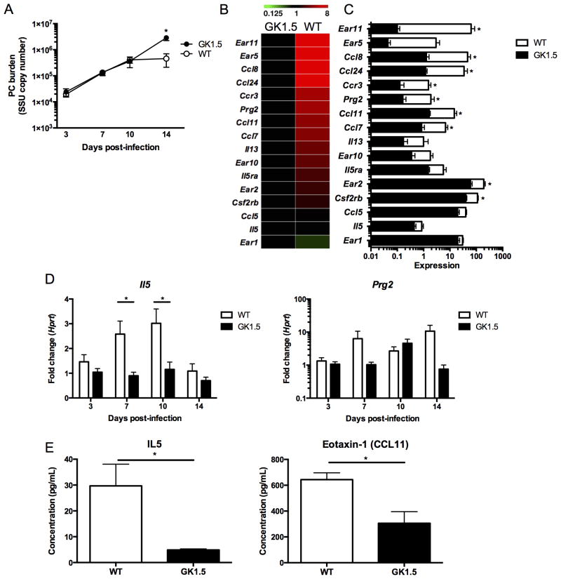
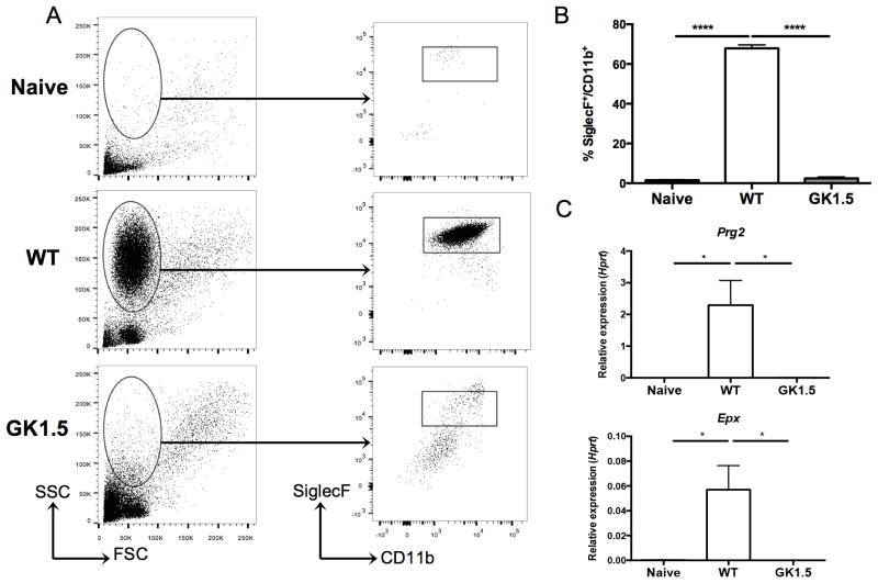
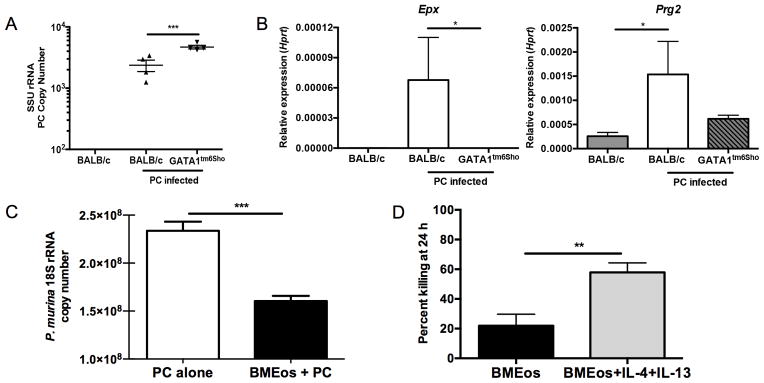
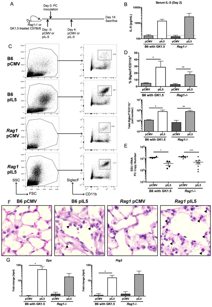
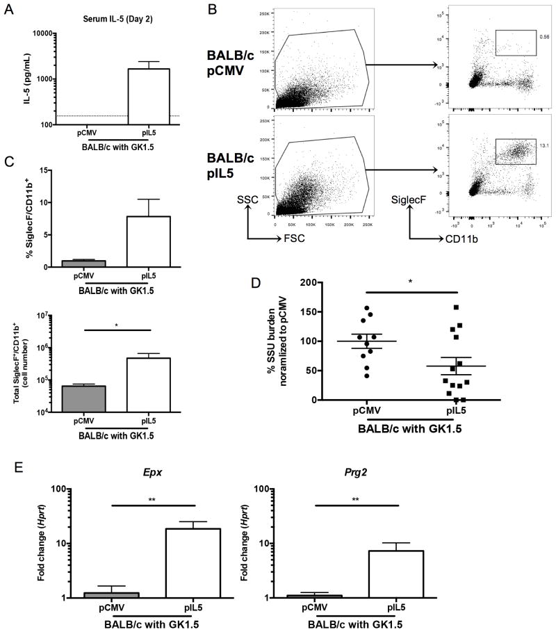
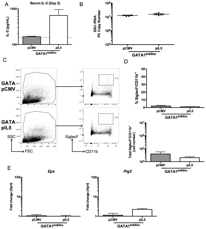
Pneumocystis pneumonia remains a common opportunistic infection in the diverse immunosuppressed population. One clear risk factor for susceptibility to Pneumocystis is a declining CD4(+) T cell count in the setting of HIV/AIDS or primary immunodeficiency. Non-HIV-infected individuals taking immunosuppressive drug regimens targeting T cell activation are also susceptible. Given the crucial role of CD4(+) T cells in host defense against Pneumocystis, we used RNA sequencing of whole lung early in infection in wild-type and CD4-depleted animals as an unbiased approach to examine mechanisms of fungal clearance. In wild-type mice, a strong eosinophil signature was observed at day 14 post Pneumocystis challenge, and eosinophils were increased in the bronchoalveolar lavage fluid of wild-type mice. Furthermore, eosinophilopoiesis-deficient Gata1(tm6Sho)/J mice were more susceptible to Pneumocystis infection when compared with BALB/c controls, and bone marrow-derived eosinophils had in vitro Pneumocystis killing activity. To drive eosinophilia in vivo, Rag1(-/-) mice were treated with a plasmid expressing IL-5 (pIL5) or an empty plasmid control via hydrodynamic injection. The pIL5-treated mice had increased serum IL-5 and eosinophilia in the lung, as well as reduced Pneumocystis burden, compared with mice treated with control plasmid. In addition, pIL5 treatment could induce eosinophilia and reduce Pneumocystis burden in CD4-depleted C57BL/6 and BALB/c mice, but not eosinophilopoiesis-deficient Gata1(tm6Sho)/J mice. Taken together, these results demonstrate that an early role of CD4(+) T cells is to recruit eosinophils to the lung and that eosinophils are a novel candidate for future therapeutic development in the treatment of Pneumocystis pneumonia in the immunosuppressed population.


Figures

Figure 1
Figure 1. RNA sequencing of whole lung shows a prominent CD4-dependent eosinophil signature at day 14 of Pneumocystis infection
A. Wild type or GK1.5 treated CD4-depleted C57Bl/6 mice were infected with 2.0 × 10⁶ cysts/ml of Pneumocystis and were sacrificed at day 3, 7, 10, or 14 following infection (n=5 at each time point). Pneumocystis burden was calculated by qRT-PCR of the small subunit ribosomal RNA and a significant decrease was seen at day 14 (p<0.01 by student’s t-test). B. RNA sequencing of whole lung RNA at day 14 in GK1.5 treated and wild type mice shows increase in expression in genes associated with eosinophils (n=4 in each group). C. Histogram of expression values from heat map in B with * indicating p<0.05 by student’s t-test. D. Il5 expression over the course of Pneumocystis infection normalized to Hprt and GK1.5 day 3 (fold change) shows increase of il5 at day 7 and 10 in wild type mice (* p<0.05, student’s t-test). Similar to the expression pattern seen by RNA sequencing, a ten-fold increase in Prg2 is seen at day 14 by qRT-PCR (p>0.05). E. IL-5 and Eotaxin-1 (CCL11) protein levels in lung homogenate at day 14 as determined by luminex (* p<0.05).
Figure 2
Figure 2. CD4-dependent recruitment of eosinophils to the lung at day 14 of Pneumocystis infection
A. Bronchoalveolar lavage (BAL) of naïve (uninfected), wild type, and GK1.5 treated CD4-depleted mice 14 days post-inoculation with Pneumocystis shows a large population of cells with high side-scatter in wild type mice (left panel). The cells were gated as shown (left panel), and a SiglecF+CD11b+ population was seen in the wild type, but not the naïve or GK1.5 treated animals (right panel). B. Significant increase in percentage of SiglecF+CD11b+ cells in wild type animals compared to naïve and GK1.5 treated animals (n=4–5, **** p<0.0001 by one-way ANOVA with Tukey’s multiple comparisons). C. qRT-PCR for Epx (top) and Prg2 (bottom) on RNA extracted from BAL cell pellets shows significant increase in expression in wild type animals compared to naïve and GK1.5 treated animals (* p<0.05 by one-way ANOVA with Tukey’s multiple comparisons).
Figure 3
Figure 3. Eosinophils contribute to control of Pneumocystis infection both in vitro and in vivo
A. BALB/c and Gata1tm6Sho/J knockout mice were infected with Pneumocystis and sacrificed at day 14 post-infection and SSU burden was quantified by qRT-PCR (** p<0.01 by student’s T test). Uninfected BALB/c mice have no detectable Pneumocystis burden. B. qRT-PCR for Epx (left) and Prg2 (right) on RNA from whole lung shows significant increase in BALB/c mice infected with Pneumocystis compared to uninfected BALB/c and infected Gata1tm6Sho/J knockout mice (* p<0.05 by Kruskal-Wallis test with Dunn’s multiple comparisons test). C. Bone marrow derived eosinophils from BALB/c mice demonstrate anti-Pneumocystis activity when co-cultured in vitro for 24 hours at an eosinophil to P. murina cyst ratio of 100:1 (*** p<0.0001, student’s t-test). D. Bone marrow derived eosinophils show enhanced killing activity when co-cultured with Pneumocystis in the presence of 10 ng/ml of IL-4 and IL-13 compared to Pneumocystis alone (** p<0.01 by student’s T test).
Figure 4
Figure 4. Treatment of CD4-depleted C57Bl/6 and Rag1−/− mice with pIL5 results in eosinophilia in whole lung and decreased Pneumocystis burden
A. Schematic showing timing of hydrodynamic injection of either pCMV or pIL5 (day −3 and 4) and Pneumocystis inoculation (day 0) into GK1.5 treated C57Bl/6 (B6) or Rag1−/− mice. B. Serum IL-5 ELISA at day 2 post infection shows log-fold increase in IL-5 in pIL5 treated animals (n=4–7, dotted line represents limit of detection). C. Digested whole lung shows a high side scatter population in the pIL5 groups compared to the pCMV treated groups (left panel). The cells were gated as shown (left panel), and a SiglecF+CD11b+ population was seen in both the B6 and Rag1−/− mice treated with pIL5 at day 14 post infection (right panel). D. pIL5 treatment resulted in a statistically significant increase in percentage (top) and total number (bottom) of SiglecF+CD11b+ cells (* p<0.05, ** p<0.01 by student’s t-test). E. pIL5 treatment results in statistically significant reduction in Pneumocystis burden as measured by qRT-PCR of small subunit ribosomal RNA at day 14 post infection (* p<0.05, *** p<0.001 by student’s t-test). F. H&E staining on paraffin-embedded lung sections demonstrating eosinophils (black arrowheads) in both the B6 and Rag1−/− mice treated with pIL5 at 60X magnification. G. qRT-PCR for Epx (left) and Prg2 (right) on RNA extracted from whole lung shows significant log-fold increase in expression in pIL5 treated animals compared to pCMV treated mice (* p<0.05 by student’s T test).
Figure 5
Figure 5. pIL5 treatment can reduce Pneumocystis burden in CD4-depleted BALB/c mice
A. Serum IL-5 ELISA at day 2 post infection shows over a log-fold increase in IL-5 in pIL5 treated BALB/c mice (n=10–13, dotted line represents limit of detection). B. Digested whole lung shows a high side scatter population in the pIL5 groups compared to the pCMV treated groups (left panel). The cells were gated as shown (left panel), and a SiglecF+CD11b+ population was seen in the BALB/c mice treated with pIL5 (right panel). C. pIL5 treatment resulted in an increase in percentage (top) and total number (bottom) of SiglecF+CD11b+ cells (* p<0.05, Mann Whitney test). D. pIL5 treatment results in reduction in Pneumocystis burden as measured by qRT-PCR of small subunit ribosomal RNA at day 14 post infection (* p=0.0481 by student’s t-test). E. qRT-PCR for Epx (left) and Prg2 (right) on RNA extracted from whole lung shows significant log-fold increase in expression in pIL5 treated animals compared to pCMV treated mice (** p<0.01 by Mann Whitney test).
Figure 6
Figure 6. pIL5 treatment cannot rescue eosinophil-deficient Gata1tm6Sho/J knockout mice
A. Serum IL-5 ELISA at day 2 post infection shows nearly a log increase in Gata1tm6Sho/J pIL5 treated animals (n=4, dotted line represents limit of detection). B. pIL5 treatment does not reduce Pneumocystis burden at day 14 post infection in Gata1tm6Sho/J mice as measured by qRT-PCR of small subunit ribosomal RNA. C. Digested whole lung shows no difference in high side scatter populations (left panel) or SiglecF+CD11b+ cells (right panel) independent of pIL5 treatment status. D. pIL5 treated Gata1tm6Sho/J mice showed no difference in percentage (top) or total number (bottom) of SiglecF+CD11b+ cells. E. qRT-PCR for Epx (left) and Prg2 (right) on RNA extracted from whole lung shows no increase in eosinophil associated genes in Gata1tm6Sho/J mice.

References

    1. Eddens T, Kolls JK. Pathological and protective immunity to Pneumocystis infection. Semin Immunopathol. 2015;37:153–162. - PMC - PubMed
    1. Antiretroviral Therapy Cohort C. Mocroft A, Sterne JA, Egger M, May M, Grabar S, Furrer H, Sabin C, Fatkenheuer G, Justice A, Reiss P, d’Arminio Monforte A, Gill J, Hogg R, Bonnet F, Kitahata M, Staszewski S, Casabona J, Harris R, Saag M. Variable impact on mortality of AIDS-defining events diagnosed during combination antiretroviral therapy: not all AIDS-defining conditions are created equal. Clin Infect Dis. 2009;48:1138–1151. - PMC - PubMed
    1. Huang L, Cattamanchi A, Davis JL, den Boon S, Kovacs J, Meshnick S, Miller RF, Walzer PD, Worodria W, Masur H I. V. a. O. P. S. International, and H. I. V. S. Lung. HIV-associated Pneumocystis pneumonia. Proc Am Thorac Soc. 2011;8:294–300. - PMC - PubMed
    1. Malin AS, Gwanzura LK, Klein S, Robertson VJ, Musvaire P, Mason PR. Pneumocystis carinii pneumonia in Zimbabwe. Lancet. 1995;346:1258–1261. - PubMed
    1. Morris A, Lundgren JD, Masur H, Walzer PD, Hanson DL, Frederick T, Huang L, Beard CB, Kaplan JE. Current epidemiology of Pneumocystis pneumonia. Emerging infectious diseases. 2004;10:1713–1720. - PMC - PubMed

Publication types

MeSH terms