. 2015 May 20;7(288):288ra79.
doi: 10.1126/scitranslmed.aaa5094.

The αvβ1 integrin plays a critical in vivo role in tissue fibrosis

Affiliations

The αvβ1 integrin plays a critical in vivo role in tissue fibrosis

Nilgun I Reed et al. Sci Transl Med. .

Abstract

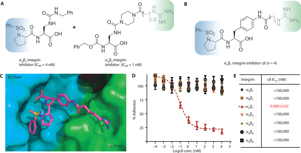
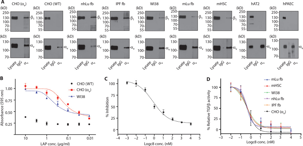
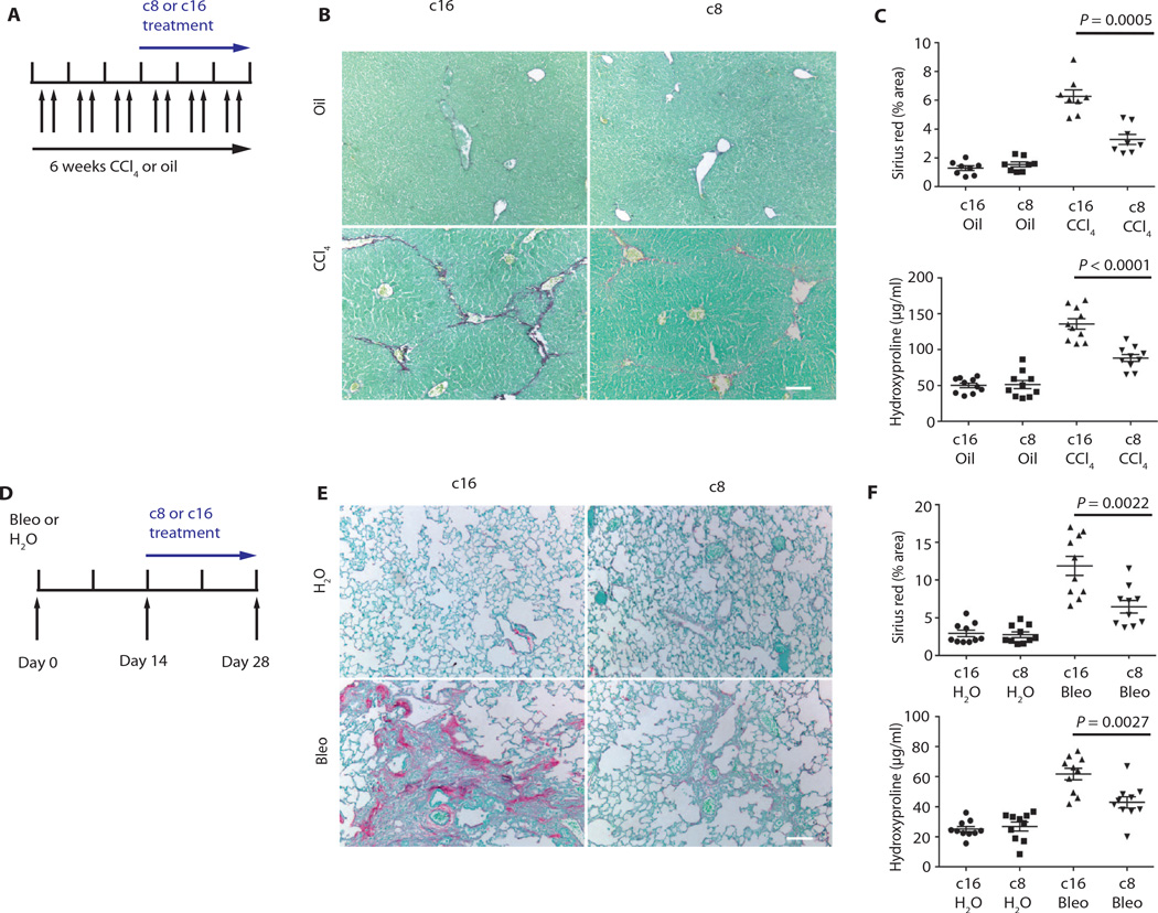
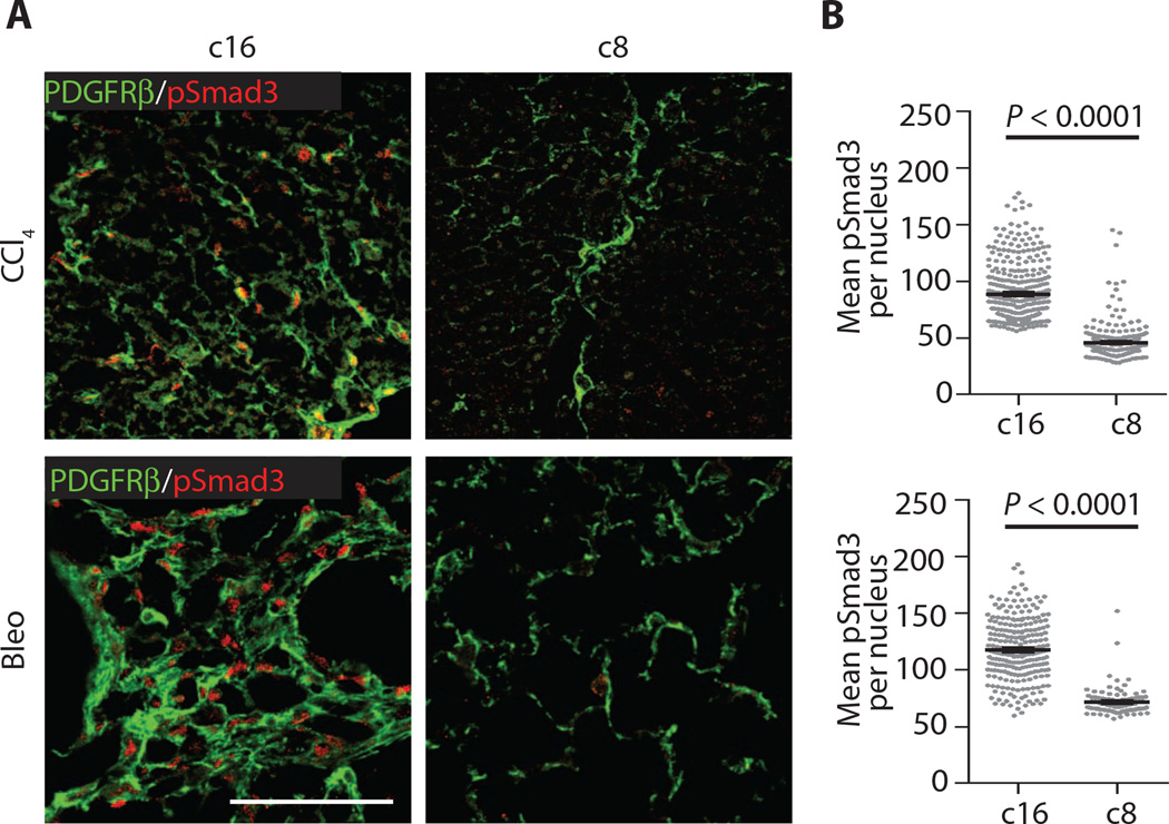
Integrins are transmembrane heterodimeric receptors that contribute to diverse biological functions and play critical roles in many human diseases. Studies using integrin subunit knockout mice and inhibitory antibodies have identified important roles for nearly every integrin heterodimer and led to the development of a number of potentially useful therapeutics. One notable exception is the αvβ1 integrin. αv and β1 subunits are individually present in numerous dimer pairs, making it challenging to infer specific roles for αvβ1 by genetic inactivation of individual subunits, and αvβ1 complex-specific blocking antibodies do not yet exist. We therefore developed a potent and highly specific small-molecule inhibitor of αvβ1 to probe the function of this understudied integrin. We found that αvβ1, which is highly expressed on activated fibroblasts, directly binds to the latency-associated peptide of transforming growth factor-β1 (TGFβ1) and mediates TGFβ1 activation. Therapeutic delivery of this αvβ1 inhibitor attenuated bleomycin-induced pulmonary fibrosis and carbon tetrachloride-induced liver fibrosis, suggesting that drugs based on this lead compound could be broadly useful for treatment of diseases characterized by excessive tissue fibrosis.


Figures

Fig. 1
Fig. 1. αvβ1 integrin inhibitor design and demonstration of potency and specificity
(A) Design principle of αvβ1 integrin inhibitor by combining a positively charged guanidine moiety in αvβ3 integrin inhibitor (blue shading) and a sulfonamidoproline moiety in α2β1 integrin inhibitor (green shading). (B) Structures of αvβ1 integrin–specific inhibitor c8. (C) Docking model of αvβ1 integrin inhibitor c8 bound to αvβ1 integrin, where α and β subunits are, respectively, in green and blue. The model predicted that a linker length of n = 3 or 4 would have the highest affinity. (D) Dose-dependent cell adhesion assay of c8 against all αv and related integrins. (E) Curve-fitted IC50 of c8 against RGD (arginine–glycine– aspartic acid)–binding integrins in cell adhesion assay. Data represent means ± SEM; n = 3 (three experimental replicates).
Fig. 2
Fig. 2. αvβ1 is expressed on pulmonary and hepatic fibroblasts and mediates adhesion to TGFβ1 LAP and activation of latent TGFβ
(A) Coimmunoprecipitation and Western blot reveal expression of αvβ1 heterodimers in human and murine cell lines from the liver and lung. Cell lysates were immunoprecipated with antibodies to αv (antibody RMV-7 for murine cells or L230 for all other cells), followed by Western blotting for either αv (lower panels, to control for capture, antibody 611012) or β1 (upper panels, antibody 04-1109). nhLu fb control (normal human lung fibroblasts from an uninjured control subject); IPF fb (lung fibroblasts isolated from a patient with IPF); mLu fb (murine lung fibroblasts); mHSC (murine hepatic stellate cells); WI38 (diploid human lung fibroblast); CHO [WT (wild-type)] control α5-deficient CHO cells (which lack expression of αvβ1); CHO (αv) (α5-deficient CHO cells engineered to express the αvβ1); hAT2 (human alveolar type 2 cells, which lack expression of αvβ1); hPAEC (human pulmonary artery endothelial cells, which lack expression of αvβ1). IgG, immunoglobulin G. (B) WT CHO cells (lacking αvβ1) adhere poorly, whereas CHO cells with forced expression of αvβ1 (CHO αv) and WI38 cells strongly adhere to TGFβ1 LAP. (C) WI38 cell adhesion to TGFβ1 LAP (0.3 µg/ml) is inhibited by c8 (IC50 = 0.72 nM). (D) c8 treatment reduced activation of TGFβ by cells expressing αvβ1 (IC50 range, 0.35 to 0.50nM). Fibroblasts (as indicated) were cocultured with TGFβ reporter (PAI1-luciferase) cell line in the presence of a range of concentrations of c8. Data represent means ± SEM, n = 3 (three experimental replicates).
Fig. 3
Fig. 3. c8 protects from liver and lung fibrosis
(A) Effects of c8 or c16 on mouse fibrosis model (liver). c8 or c16 (inactive control compound) was continuously delivered by Alzet pump beginning 3 weeks after intraperitoneal administration with oil (sham) or CCl4 to induce liver fibrosis. (B and C) Treatment with c8 significantly reduced liver fibrosis, as determined by (B) Sirius red staining (collagen deposition) of liver tissue after olive oil (top panels) or CCl4 treatment (bottom panels), (C) digital image analysis quantification of collagen staining, and hydroxyproline analysis. Sirius red (liver) n = 8; P = 0.0005. Hydroxyproline (liver) n = 10; P < 0.0001. (D) Effects of c8 or c16 on murine fibrosis model (lung). c8 or c16 was continuously delivered to mice using Alzet pumps beginning 14 days after intratracheal instillation of bleomycin (Bleo) to induce pulmonary fibrosis or water (H2O). (E and F) Treatment with c8 significantly reduced lung fibrosis, as determined by (E) Sirius red staining (collagen deposition) of lung tissue after water (top panels) or bleomycin treatment (bottom panels), (F) digital image analysis quantification of collagen staining, and hydroxyproline analysis. Sirius red (lung) n = 10; P = 0.0022. Hydroxyproline (lung) n = 10; P = 0.0027. Data represent means ± SEM. Scale bars, 100 µm. P values were calculated using the unpaired Student’s t tests.
Fig. 4
Fig. 4. Reduced pSmad3 in mice treated with c8
(A) Representative liver (top panels) and lung (bottom panels) sections from mice treated with c16 or c8 after induced fibrotic injury stained for fibroblasts [PDGFRβ (plateletderived growth factor receptor β), green] and pSmad3 (red). (B) Quantification of pSmad3 nuclear intensity within individual PDGFRβ+ cells documents a significant reduction in fibroblast-specific pSmad3 in fibrotic mice treated with c8. Data represent means ± SEM; n = 102 (Bleo c8), n = 254 (Bleo c16), n = 262 (CCl4 c8), and n = 361(CCl4 c16). For both comparisons, P < 0.0001 (shown is a representative example of the distribution of individual pSmad3 mean fluorescence intensities in PDGFRβ+ cells, and the average of these means, for a single sample condition). Scale bar, 100 µm. P values were calculated using the unpaired Student’s t tests.

Comment in

References

    1. Hynes RO. Integrins: Versatility, modulation, and signaling in cell adhesion. Cell. 1992;69:11–25. - PubMed
    1. Munger JS, Sheppard D. Cross talk among TGF-β signaling pathways, integrins, and the extracellular matrix. Cold Spring Harb. Perspect. Biol. 2011;3:a005017. - PMC - PubMed
    1. Hynes RO. Integrins: Bidirectional, allosteric signaling machines. Cell. 2002;110:673–687. - PubMed
    1. Sheppard D. In vivo functions of integrins: Lessons from null mutations in mice. Matrix Biol. 2000;19:203–209. - PubMed
    1. Chen C, Sheppard D. Identification and molecular characterization of multiple phenotypes in integrin knockout mice. Methods Enzymol. 2007;426:291–305. - PubMed

Publication types

MeSH terms