. 2015 Jul;8(4):757-65.
doi: 10.1161/CIRCHEARTFAILURE.115.002210. Epub 2015 May 20.

Effects of Intracoronary Infusion of Escalating Doses of Cardiac Stem Cells in Rats With Acute Myocardial Infarction

Affiliations

Effects of Intracoronary Infusion of Escalating Doses of Cardiac Stem Cells in Rats With Acute Myocardial Infarction

Xian-Liang Tang et al. Circ Heart Fail. 2015 Jul.

Abstract

Background: Although c-kit(pos) cardiac stem cells (CSCs) preserve left ventricular (LV) function and structure after myocardial infarction, CSC doses have been chosen arbitrarily, and the dose-effect relationship is unknown.

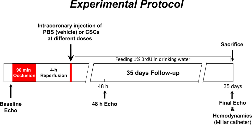
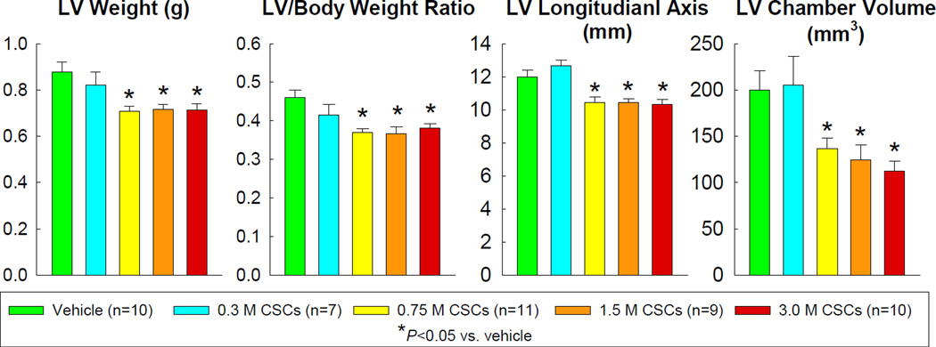
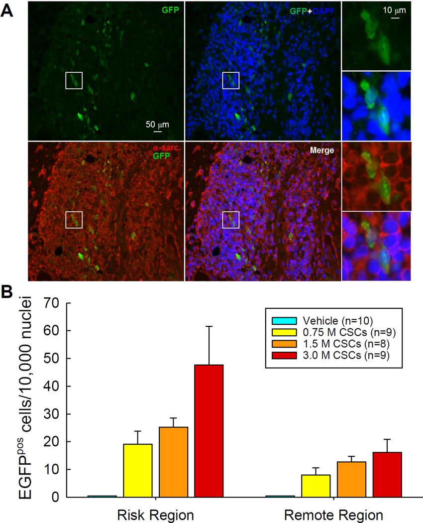
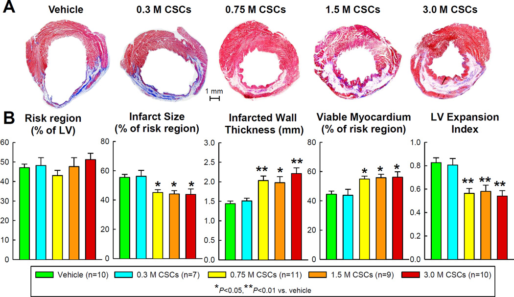
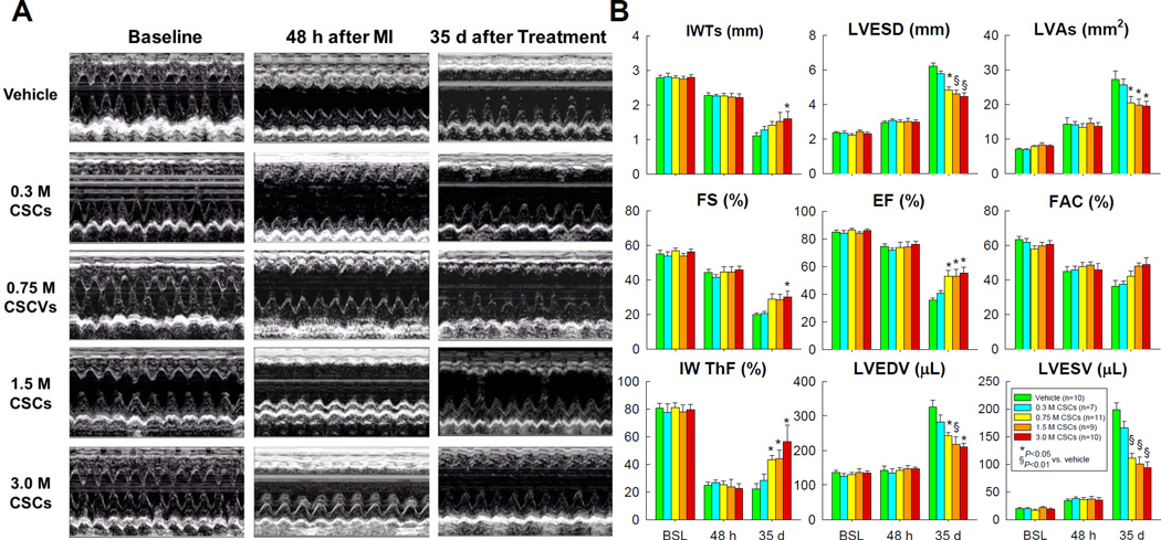
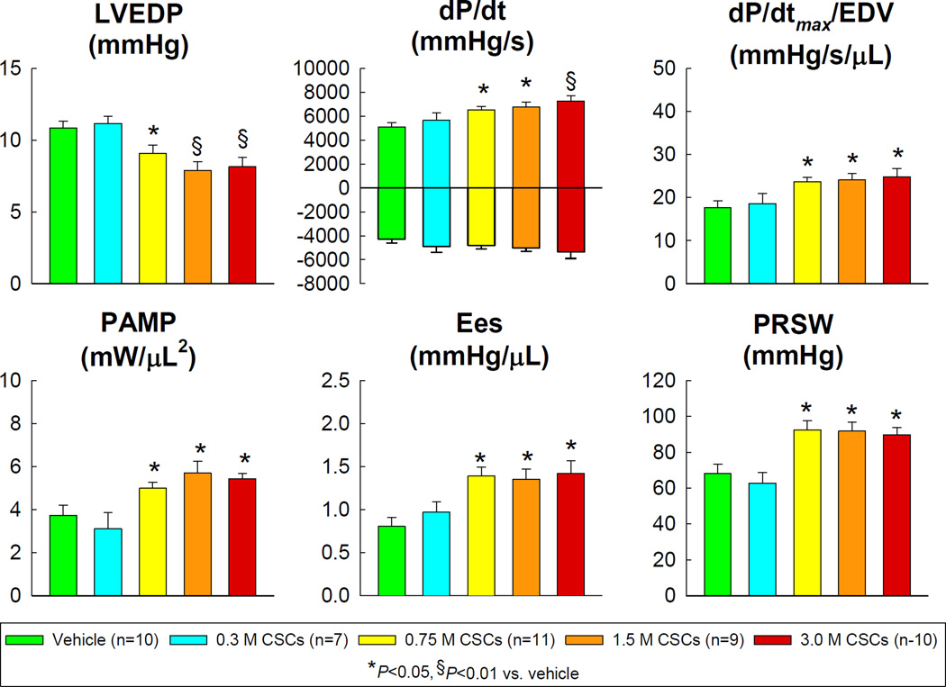
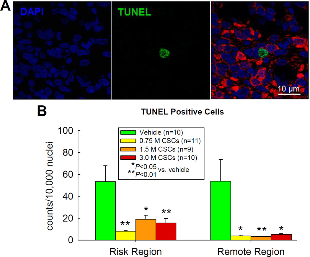
Methods and results: Rats underwent a 90-minute coronary occlusion followed by 35 days of reperfusion. Vehicle or CSCs at 5 escalating doses (0.3×10(6), 0.75×10(6), 1.5×10(6), 3.0×10(6), and 6.0×10(6) cells/heart) were given intracoronarily 4 h after reperfusion. The lowest dose (0.3×10(6)) had no effect on LV function and morphology, whereas 0.75, 1.5, and 3.0×10(6) significantly improved regional and global LV function (echocardiography and hemodynamic studies). These 3 doses had similar effects on echocardiographic parameters (infarct wall thickening fraction, LV end-systolic and end-diastolic volumes, LV ejection fraction) and hemodynamic variables (LV end-diastolic pressure, LV dP/dtmax, preload adjusted maximal power, end-systolic elastance, preload recruitable stroke work) and produced similar reductions in apoptosis, scar size, infarct wall thinning, and LV expansion index and similar increases in viable myocardium in the risk region (morphometry). Infusion of 6.0×10(6) CSCs markedly increased postprocedural mortality. Green fluorescent protein and 5-bromo-2'-deoxyuridine staining indicated that persistence of donor cells and formation of new myocytes were negligible with all doses.

Conclusions: Surprisingly, in this rat model of acute myocardial infarction, the dose-response relationship for intracoronary CSCs is flat. A minimal dose between 0.3 and 0.75×10(6) is necessary for efficacy; above this threshold, a 4-fold increase in cell number does not produce greater improvement in LV function or structure. Further increases in cell dose are harmful.

Keywords: left ventricular function; myocardial infarction; myocardial ischemia; myocardial regeneration; progenitor cells.


Figures

Figure 1
Figure 1. Experimental protocol
Fischer 344 female rats (age 3–4 months) GFP-labeled syngeneic CSCs were injected into the aortic root during two 20-s occlusions of the ascending aorta and pulmonary artery, 10 min apart , and a 0.5 ml of volume was delivered each injection (CSCs were suspended in a total of 1.0 ml medium for each rat)
Figure 2
Figure 2
Postmortem gross measurements were conducted in perfused hearts arrested in diastole. Data are means ± SEM.
Figure 3
Figure 3. Myocardial engraftment of transplanted CSCs
(A). Representative images showing GFPpos cells in the border zone of the LV section from a rat that received 3.0 ×10⁶ CSCs. (B). Quantitative analysis was performed in the hearts with detectable GFPpos cells. Data are means ± SEM.
Figure 4
Figure 4. Morphometric analysis
(A). Representative Masson’s trichrome-stained LV sections from each of the six groups. Scar tissue and viable myocardium are identified in blue/white and red, respectively. (B). Quantitative analysis of LV morphometric parameters. The thickness of the infarcted wall was determined by averaging five measurements of LV wall thickness equally distributed within the infarcted LV region; risk region was defined as the LV area between the two edges of the infarct scar; and LV expansion index was calculated as (LV endocardial circumference/LV epicardial circumference) × (non-infarcted region wall thickness/risk region wall thickness). Data are means ± SEM.
Figure 5
Figure 5. Echocardiographic assessment of LV function
(A) Representative M-mode images from the vehicle, 0.3 ×10⁶, 0.75 ×10⁶, 1.5 ×10⁶, and 3.0 ×10⁶, groups recorded at baseline (before MI), 48 h after MI, and 35 d after vehicle or CSC treatment. (B). Quantitative analyses of echocardiographic parameters at baseline (BSL), 48 h after MI (48 h), and 35 d follow-up (35 d). IWTs, systolic infarcted wall thickness; LVESD, left ventricular end-systolic diameter; LVAs, left ventricular end-systolic area; FS, fractional shortening; EF, ejection fraction; FAC, fractional area change; IW ThF, infarcted wall thickening fraction; LVEDV, left ventricular end-diastolic volume; and LVESV, left ventricular end-systolic volume. Data are means ± SEM.
Figure 6
Figure 6
Hemodynamic assessment of LV function at 35 d follow-up. LVEDP, left ventricular end-diastolic pressure; PAMP, preload adjusted maximal power; Ees, end-systolic elastance; and PRSW, preload recruitable stroke work. Data are means ± SEM.
Figure 7
Figure 7. Analysis of apoptosis by TUNEL staining
(A) Representative confocal microscopic images in the border zone of a 3.0 ×10⁶ CSC-treated rat showing TUNEL positive nuclei (green), DAPI staining (blue), and a merge of TUNEL and DAPI staining; (B) Quantitative analysis of TUNEL positive nuclei in the risk and remote (noninfarcted) region in the vehicle- and three doses of CSC-treated groups. Data are means ± SEM.
Figure 8
Figure 8. Proliferation of transplanted CSCs
(A) Representative confocal microscopic images from a 3.0 ×10⁶ CSC-treated rat showing BrdU uptake in the infarcted region during the 35 d follow-up. Yellow arrows indicate GFPpos cells that are BrdU positive. (B) Quantitative analysis of BrdUpos cells, BrdUpos/α-sarcpos cells (cells with colocalization of BrdU [FITC] and α-sarcomeric actin [TRITC]), and BrdUpos/α-sarcpos/GFPpos cells (cells with colocalization of BrdU [FITC], α-sarcomeric actin [TRITC], and GFP [TRITC]) in the risk and remote (noninfarcted) regions. Data are means ± SEM.

References

    1. Beltrami AP, Barlucchi L, Torella D, Baker M, Limana F, Chimenti S, Kasahara H, Rota M, Musso E, Urbanek K, Leri A, Kajstura J, Nadal-Ginard B, Anversa P. Adult cardiac stem cells are multipotent and support myocardial regeneration. Cell. 2003;114:763–776. - PubMed
    1. Fischer KM, Cottage CT, Wu W, Din S, Gude NA, Avitabile D, Quijada P, Collins BL, Fransioli J, Sussman MA. Enhancement of myocardial regeneration through genetic engineering of cardiac progenitor cells expressing pim-1 kinase. Circulation. 2009;120:2077–2087. - PMC - PubMed
    1. Oskouei BN, Lamirault G, Joseph C, Treuer AV, Landa S, Da Silva J, Hatzistergos K, Dauer M, Balkan W, McNiece I, Hare JM. Increased potency of cardiac stem cells compared with bone marrow mesenchymal stem cells in cardiac repair. Stem Cells Transl Med. 2012;1:116–124. - PMC - PubMed
    1. Bearzi C, Rota M, Hosoda T, Tillmanns J, Nascimbene A, De Angelis A, Yasuzawa-Amano S, Trofimova I, Siggins RW, Lecapitaine N, Cascapera S, Beltrami AP, D'Alessandro DA, Zias E, Quaini F, Urbanek K, Michler RE, Bolli R, Kajstura J, Leri A, Anversa P. Human cardiac stem cells. Proc Natl Acad Sci U S A. 2007;104:14068–14073. - PMC - PubMed
    1. Duran JM, Makarewich CA, Sharp TE, Starosta T, Zhu F, Hoffman NE, Chiba Y, Madesh M, Berretta RM, Kubo H, Houser SR. Bone-derived stem cells repair the heart after myocardial infarction through transdifferentiation and paracrine signaling mechanisms. Circ Res. 2013;113:539–552. - PMC - PubMed

Publication types

MeSH terms