. 2015 Aug;89(15):7776-85.
doi: 10.1128/JVI.00467-15. Epub 2015 May 20.

Conservation of G-Protein Epitopes in Respiratory Syncytial Virus (Group A) Despite Broad Genetic Diversity: Is Antibody Selection Involved in Virus Evolution?

Affiliations

Conservation of G-Protein Epitopes in Respiratory Syncytial Virus (Group A) Despite Broad Genetic Diversity: Is Antibody Selection Involved in Virus Evolution?

Alfonsina Trento et al. J Virol. 2015 Aug.

Abstract

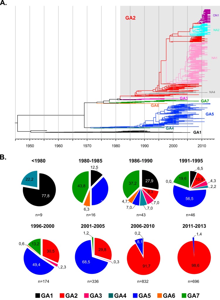
Worldwide G-glycoprotein phylogeny of human respiratory syncytial virus (hRSV) group A sequences revealed diversification in major clades and genotypes over more than 50 years of recorded history. Multiple genotypes cocirculated during prolonged periods of time, but recent dominance of the GA2 genotype was noticed in several studies, and it is highlighted here with sequences from viruses circulating recently in Spain and Panama. Reactivity of group A viruses with monoclonal antibodies (MAbs) that recognize strain-variable epitopes of the G glycoprotein failed to correlate genotype diversification with antibody reactivity. Additionally, no clear correlation was found between changes in strain-variable epitopes and predicted sites of positive selection, despite both traits being associated with the C-terminal third of the G glycoprotein. Hence, our data do not lend support to the proposed antibody-driven selection of variants as a major determinant of hRSV evolution. Other alternative mechanisms are considered to account for the high degree of hRSV G-protein variability.

Importance: An unusual characteristic of the G glycoprotein of human respiratory syncytial virus (hRSV) is the accumulation of nonsynonymous (N) changes at higher rates than synonymous (S) changes, reaching dN/dS values at certain sites predictive of positive selection. Since these sites cluster preferentially in the C-terminal third of the G protein, like certain epitopes recognized by murine antibodies, it was proposed that immune (antibody) selection might be driving the apparent positive selection, analogous to the antigenic drift observed in the influenza virus hemagglutinin (HA). However, careful antigenic and genetic comparison of the G glycoprotein does not provide evidence of antigenic drift in the G molecule, in agreement with recently published data which did not indicate antigenic drift in the G protein with human sera. Alternative explanations to the immune-driven selection hypothesis are offered to account for the high level of G-protein genetic diversity highlighted in this study.


Figures

FIG 1
FIG 1
Phylogeny of hRSV group A viruses and genotype temporal dominance. (A) Maximum clade credibility (MCC) tree from Bayesian analysis of 1,485 unique nucleotide sequences of the G-protein gene ectodomain of hRSV group A retrieved from GenBank. Clades are colored according the genotype classification shown in Table 1. (B) Frequencies of the different genotypes in 5-year periods from 1956 to 2013. The number of sequences (n) included in each period is indicated below the charts.
FIG 2
FIG 2
Phylogenetic tree of hRSV group A sequences from recent epidemics in Madrid and Panama. The maximum-likelihood phylogenetic tree was constructed on the basis of nucleotide sequences of the G-protein ectodomain obtained from Madrid (diamonds) and Panama (circles) samples. Virus nomenclature follows the general consensus, with the last two digits referring to year of isolation. The bar represents 0.02 nucleotide substitution per site, and the tree is unrooted. Numbers at the internal nodes represent the bootstrap probabilities (1,000 replicates). Only bootstrap values of >70 are shown. The number of sequences identical to those shown in the figure is indicated in parentheses at right of the sample name. Asterisks denote viruses included in the analysis shown in Fig. 3 and 4.
FIG 3
FIG 3
Reactivity of group A viruses with MAbs. Sequences of the viral strains used in this experiment were used to build the phylogenetic tree shown on the left, as for Fig. 2. Each virus was used to infect HEp-2 cell cultures, which were stained at 24 h after infection with the indicated MAbs and anti-mouse fluorescein-linked antibody (GE Healthcare). Two panels of strain-variable MAbs were used: one panel included MAbs 63G, 25G, 78G, and 68G, obtained from mice inoculated with the Long strain of hRSV (17), and the other included MAbs 021/12G, 021/10G, 021/9G, 021/8G, 021/7G, and 021/16G, obtained from mice inoculated with Mon/3/88 virus (18). MAb 021/1G, which recognizes a conserved epitope of hRSV G protein, was included as control. Numbers shown within the boxes are the fluorescence values after normalization, so that fluorescence of Long with each Long-specific MAb was normalized to 100% and similarly for the Mon/3/88 virus with the MAbs specific for this virus. Squares with fluorescence values of >50% have a black background, those with values between 25% and 50% have a gray background, and those with values of <25% have a white background. The results are representative of five independent determinations.
FIG 4
FIG 4
UV light photographs of cultures used for the quantitative analysis in Fig. 3.
FIG 5
FIG 5
Sequence alignment of the C-terminal thirds of G-protein sequences. (A) Alignment of partial (C-terminal) G-protein sequences of the viruses used for Fig. 3. Numbering is shown above the Long sequence, which is used as a reference for the next two viruses. The entire sequence of Mon/3/88 is also shown as a reference for the rest of viruses. Only the amino acid changes are indicated. A lack of change is denoted by a dot. Asterisks indicate stop codons. Note the 72-nt duplicated sequence in four viruses, which forces the gaps denoted by hyphens in the other sequences. Residues that showed changes in mutants selected with the indicated MAbs are indicated by arrows at the top. (B) Sites of positive selection predicted in the indicated studies (11, 13, 30, 31, 33, 49–53) are denoted by small circles below the corresponding amino acid.

References

    1. Nair H, Nokes DJ, Gessner BD, Dherani M, Madhi SA, Singleton RJ, O'Brien KL, Roca A, Wright PF, Bruce N, Chandran A, Theodoratou E, Sutanto A, Sedyaningsih ER, Ngama M, Munywoki PK, Kartasasmita C, Simoes EA, Rudan I, Weber MW, Campbell H. 2010. Global burden of acute lower respiratory infections due to respiratory syncytial virus in young children: a systematic review and meta-analysis. Lancet 375:1545–1555. doi:10.1016/S0140-6736(10)60206-1. - DOI - PMC - PubMed
    1. Falsey AR, Hennessey PA, Formica MA, Cox C, Walsh EE. 2005. Respiratory syncytial virus infection in elderly and high-risk adults. N Engl J Med 352:1749–1759. doi:10.1056/NEJMoa043951. - DOI - PubMed
    1. Collins PL, Melero JA. 2011. Progress in understanding and controlling respiratory syncytial virus: still crazy after all these years. Virus Res 162:80–99. doi:10.1016/j.virusres.2011.09.020. - DOI - PMC - PubMed
    1. Levine S, Klaiber-Franco R, Paradiso PR. 1987. Demonstration that glycoprotein G is the attachment protein of respiratory syncytial virus. J Gen Virol 68:2521–2524. doi:10.1099/0022-1317-68-9-2521. - DOI - PubMed
    1. Walsh EE, Hruska J. 1983. Monoclonal antibodies to respiratory syncytial virus proteins: identification of the fusion protein. J Virol 47:171–177. - PMC - PubMed

Publication types

MeSH terms

Associated data