. 2015 Aug;89(15):7813-28.
doi: 10.1128/JVI.00783-15. Epub 2015 May 20.

Inhibitory Effect of Individual or Combinations of Broadly Neutralizing Antibodies and Antiviral Reagents against Cell-Free and Cell-to-Cell HIV-1 Transmission

Affiliations

Inhibitory Effect of Individual or Combinations of Broadly Neutralizing Antibodies and Antiviral Reagents against Cell-Free and Cell-to-Cell HIV-1 Transmission

Randi B Gombos et al. J Virol. 2015 Aug.

Abstract

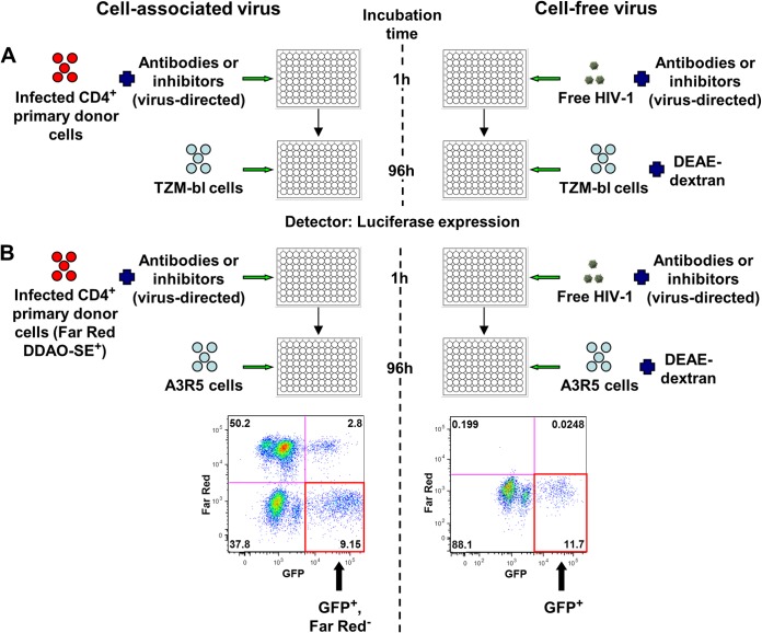
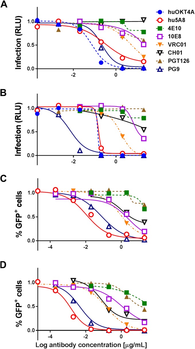
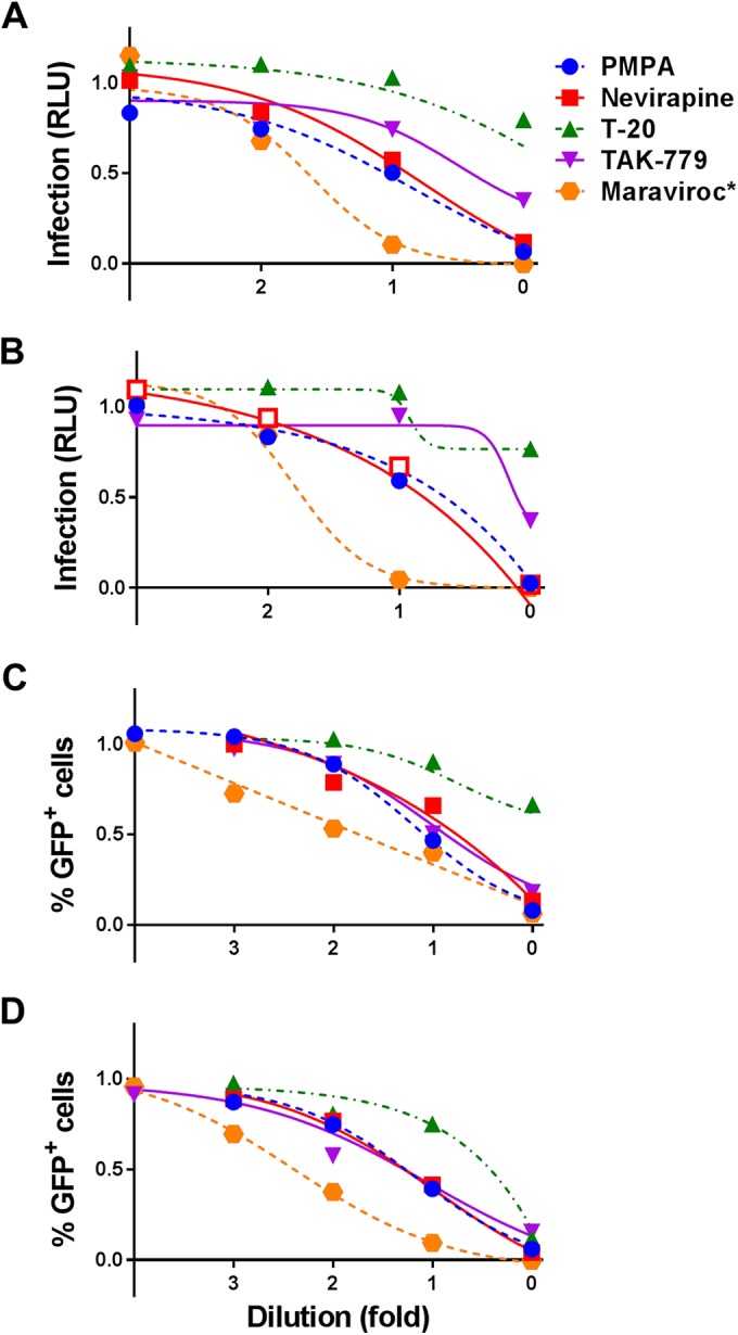
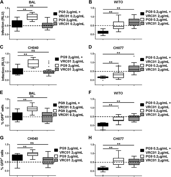
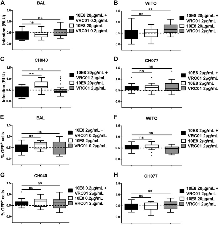
To date, most therapeutic and vaccine candidates for human immunodeficiency virus type 1 (HIV-1) are evaluated preclinically for efficacy against cell-free viral challenges. However, cell-associated HIV-1 is suggested to be a major contributor to sexual transmission by mucosal routes. To determine if neutralizing antibodies or inhibitors block cell-free and cell-associated virus transmission of diverse HIV-1 strains with different efficiencies, we tested 12 different antibodies and five inhibitors against four green fluorescent protein (GFP)-labeled HIV-1 envelope (Env) variants from transmitted/founder (T/F) or chronic infection isolates. We evaluated antibody/inhibitor-mediated virus neutralization using either TZM-bl target cells, in which infectivity was determined by virus-driven luciferase expression, or A3R5 lymphoblastoid target cells, in which infectivity was evaluated by GFP expression. In both the TZM-bl and A3R5 assays, cell-free virus or infected CD4+ lymphocytes were used as targets for neutralization. We further hypothesized that the combined use of specific neutralizing antibodies targeting HIV-1 Env would more effectively prevent cell-associated virus transmission than the use of individual antibodies. The tested antibody combinations included two gp120-directed antibodies, VRC01 and PG9, or VRC01 with the gp41-directed antibody 10E8. Our results demonstrated that cell-associated virus was less sensitive to neutralizing antibodies and inhibitors, particularly using the A3R5 neutralization assay, and the potencies of these neutralizing agents differed among Env variants. A combination of different neutralizing antibodies that target specific sites on gp120 led to a significant reduction in cell-associated virus transmission. These assays will help identify ideal combinations of broadly neutralizing antibodies to use for passive preventive antibody administration and further characterize targets for the most effective neutralizing antibodies/inhibitors.

Importance: Prevention of the transmission of human immunodeficiency virus type 1 (HIV-1) remains a prominent goal of HIV research. The relative contribution of HIV-1 within an infected cell versus cell-free HIV-1 to virus transmission remains debated. It has been suggested that cell-associated virus is more efficient at transmitting HIV-1 and more difficult to neutralize than cell-free virus. Several broadly neutralizing antibodies and retroviral inhibitors are currently being studied as potential therapies against HIV-1 transmission. The present study demonstrates a decrease in neutralizing antibody and inhibitor efficiencies against cell-associated compared to cell-free HIV-1 transmission among different strains of HIV-1. We also observed a significant reduction in virus transmission using a combination of two different neutralizing antibodies that target specific sites on the outermost region of HIV-1, the virus envelope. Therefore, our findings support the use of antibody combinations against both cell-free and cell-associated virus in future candidate therapy regimens.


Figures

FIG 1
FIG 1
Neutralization assays. (A) Frozen CD4+ cells were thawed, stimulated overnight with IL-2, and infected with one of the four GFP-labeled HIV-1 Env variants to prepare cell-associated virus. Cell-free HIV-1 or HIV-1-infected donor cells were incubated with virus-directed antibodies or inhibitors for 1 h and then added to uninfected TZM-bl target cells. DEAE-dextran was used for cell-free virus infection but not for cell-associated infection. After 96 h, Steady-Glo luciferase expression was used to quantify the amount of infection of TZM-bl cells. (B) Cell-free HIV-1 or HIV-1-infected donor cells stained with Far Red DDAO-SE were incubated with virus-directed antibodies or inhibitors for 1 h and then added to uninfected A3R5 target cells. DEAE-dextran was used for cell-free virus infection only. Flow cytometry was performed to determine the number of newly infected target cells from cell-associated (GFP+ Far Red) or cell-free (GFP+) virus. FITC, fluorescein isothiocyanate.
FIG 2
FIG 2
Infection with cell-associated or cell-free WITO ± DEAE-dextran with different antibodies. TZM-bl (A and B) or A3R5 (C and D) target cells were infected with cell-associated (A and C) or cell-free (B and D) WITO virus in the presence of different concentrations (20 to 0.00002 μg/ml) of antibodies, including anti-Flu (Ab82), A32, 4E10, 10E8, VRC01, CH01, PGT126, PG9, CH58, CH59, and the CD4-directed antibodies hu5A8 and huOKT4A. Background (negative control) was subtracted from results, and results were normalized to the no-antibody positive control and summarized as infection based on RLU (A and B) or percentage of GFP+ (Far Red) cells (C and D), where 1 is equal to 100% infection. Results are expressed as dose-response curves illustrating the median at each log dose.
FIG 3
FIG 3
Infection with cell-associated or cell-free WITO ± DEAE-dextran with different inhibitors. TZM-bl (A and B) or A3R5 (C and D) target cells were infected with cell-associated (A and C) or cell-free (B and D) WITO virus in the presence of different concentrations of inhibitors. All inhibitors were serially diluted 10-fold at various ranges of concentration (PMPA, 0.1 to 100 μM; nevirapine, 0.4 to 400 nM; T-20, 0.5 to 500 nM; TAK-779, 1 to 1,000 nM), with the exception of maraviroc, which was serially diluted 20-fold (*, 0.015625 to 2,500 nM). Background was subtracted from results, and results were normalized to the DMSO-positive control and summarized as infection based on RLU (A and B) or percentage of GFP+ (Far Red) cells (C and D), where 1 is equal to 100% infection. Results are expressed as dose-response curves illustrating the median at each dose dilution.
FIG 4
FIG 4
Neutralization of cell-associated virus by combination of two gp120-specific broadly neutralizing antibodies. Two HIV-1-directed antibodies, PG9 and VRC01, were combined at concentrations that have a partial effect to no effect on cell-associated virus infection when used individually. Infection was evaluated in TZM-bl (A to D) or A3R5 (E to H) target cells with cell-associated HIV-1 BAL (A and E), WITO (B and F), CH040 (C and G), and CH077 (D and H). Infection was measured by quantifying the RLU of TZM-bl cells (A to D) or the percentage of GFP+ (Far Red) A3R5 cells (E to H) for each cell-associated Env variant in the presence of the indicated concentrations of antibodies, used in combination or alone, where 1 is equal to 100% infection. Results were normalized and expressed as box-and-whisker plots illustrating the median, first and third quartiles, and range with outliers (solid circles). Significant differences were assessed with exact Wilcoxon rank sum tests and Holm's adjustment to compare each combination of antibodies to the single-antibody treatment. A significant difference is represented by ** (P < 0.005), and a nonsignificant difference is represented by “ns.”
FIG 5
FIG 5
Neutralization of cell-associated virus by combination of gp120- and gp41-specific broadly neutralizing antibodies. Two HIV-1-directed antibodies, 10E8 and VRC01, were combined at concentrations that have a partial effect to no effect on cell-associated virus infection when used individually. Infection was evaluated in TZM-bl (A to D) or A3R5 (E to H) target cells with cell-associated HIV-1 BAL (A and E), WITO (B and F), CH040 (C and G), and CH077 (D and H). Infection was measured by quantifying RLU of TZM-bl cells (A to D) or the percentage of GFP+ (Far Red) A3R5 cells (E to H) for each cell-associated Env variant in the presence of the indicated concentrations of antibodies, used in combination or alone, where 1 is equal to 100% infection. Results were normalized and expressed as in Fig. 4. Significant differences were assessed with exact Wilcoxon rank sum tests and Holm's adjustment to compare each combination of antibodies to the single-antibody treatment. A significant difference is represented by ** (P < 0.0075), and a nonsignificant difference is represented by “ns.”

Similar articles

Cited by

References

    1. Virgin HW, Walker BD. 2010. Immunology and the elusive AIDS vaccine. Nature 464:224–231. doi:10.1038/nature08898. - DOI - PubMed
    1. Anderson DJ, Yunis EJ. 1983. “Trojan Horse” leukocytes in AIDS. N Engl J Med 309:984–985. - PubMed
    1. Kolodkin-Gal D, Hulot SL, Korioth-Schmitz B, Gombos RB, Zheng Y, Owuor J, Lifton MA, Ayeni C, Najarian RM, Yeh WW, Asmal M, Zamir G, Letvin NL. 2013. Efficiency of cell-free and cell-associated virus in mucosal transmission of human immunodeficiency virus type 1 and simian immunodeficiency virus. J Virol 87:13589–13597. doi:10.1128/JVI.03108-12. - DOI - PMC - PubMed
    1. Salle B, Brochard P, Bourry O, Mannioui A, Andrieu T, Prevot S, Dejucq-Rainsford N, Dereuddre-Bosquet N, Le Grand R. 2010. Infection of macaques after vaginal exposure to cell-associated simian immunodeficiency virus. J Infect Dis 202:337–344. doi:10.1086/653619. - DOI - PubMed
    1. Bernard-Stoecklin S, Gommet C, Corneau AB, Guenounou S, Torres C, Dejucq-Rainsford N, Cosma A, Dereuddre-Bosquet N, Le Grand R. 2013. Semen CD4+ T cells and macrophages are productively infected at all stages of SIV infection in macaques. PLoS Pathog 9:e1003810. doi:10.1371/journal.ppat.1003810. - DOI - PMC - PubMed

Publication types

MeSH terms