. 2015 Jun 9;112(23):7231-6.
doi: 10.1073/pnas.1501327112. Epub 2015 May 20.

Circadian control of innate immunity in macrophages by miR-155 targeting Bmal1

Affiliations

Circadian control of innate immunity in macrophages by miR-155 targeting Bmal1

Anne M Curtis et al. Proc Natl Acad Sci U S A. .

Abstract

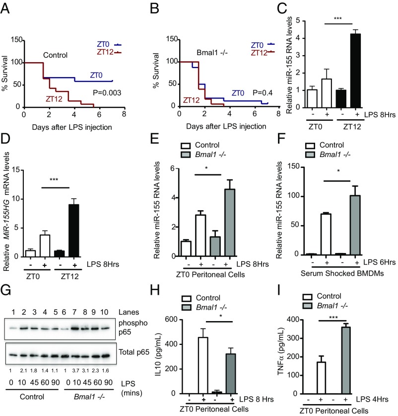
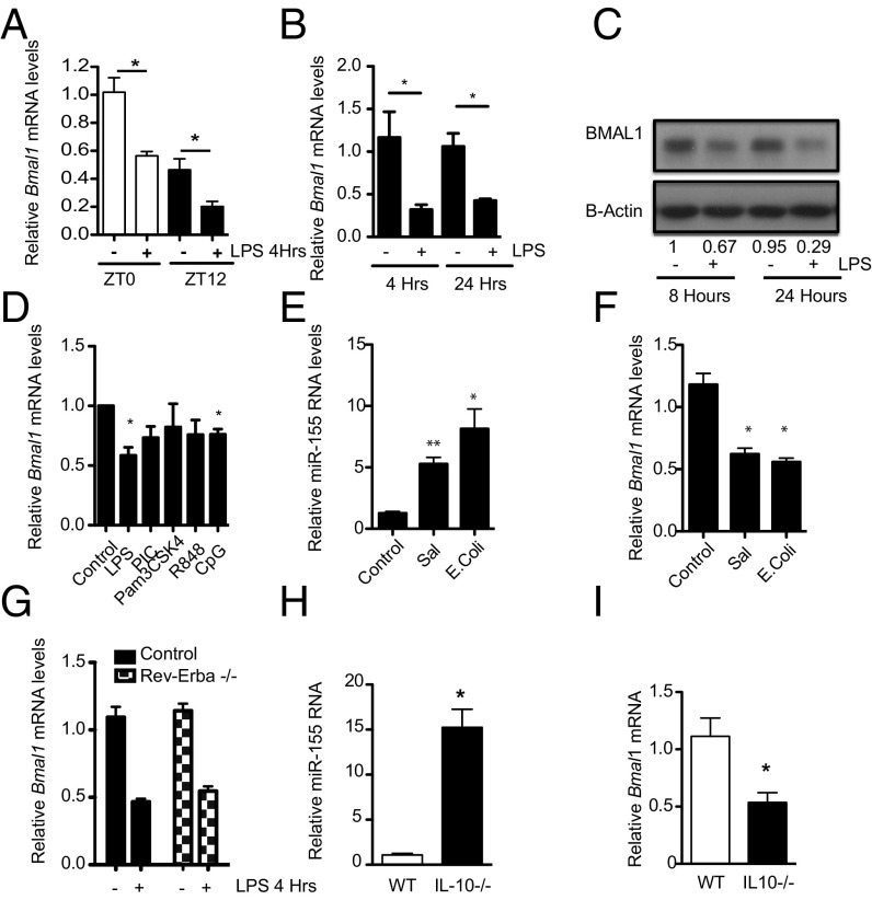
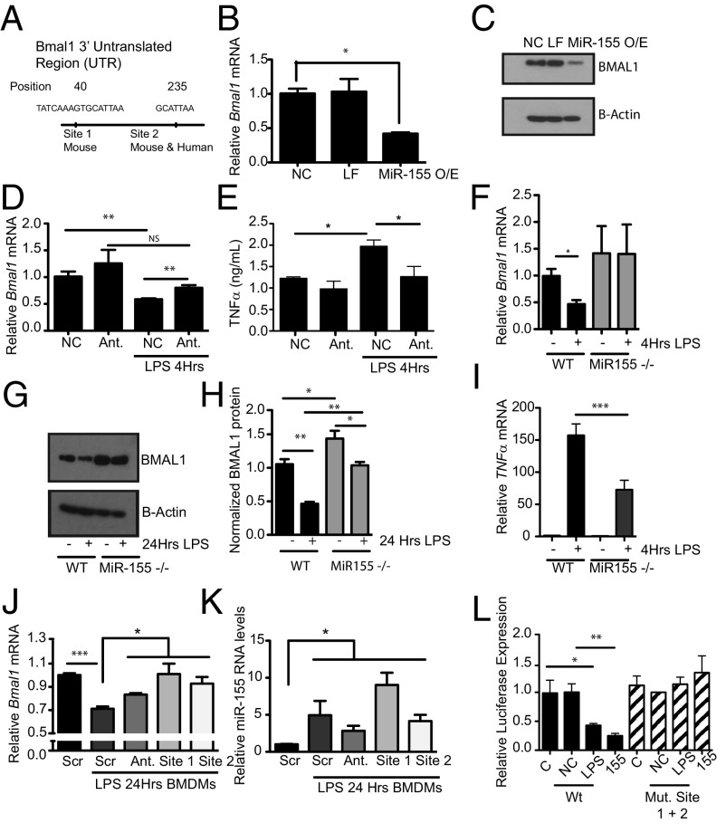
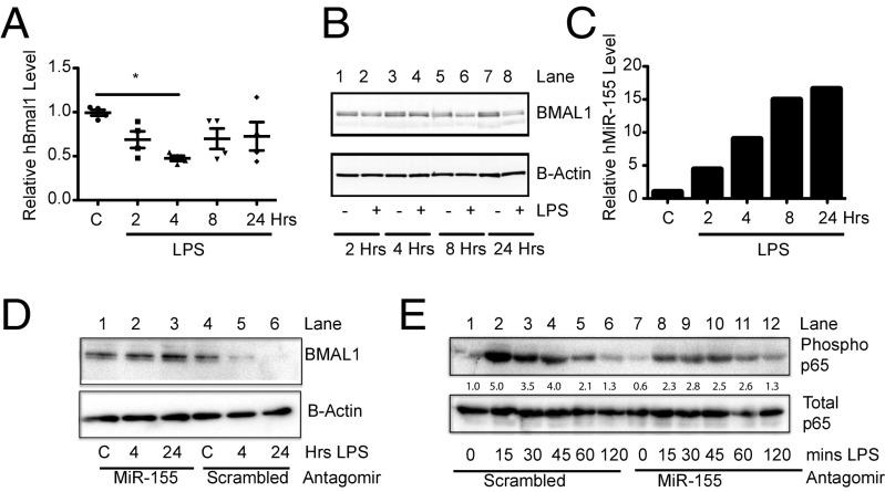
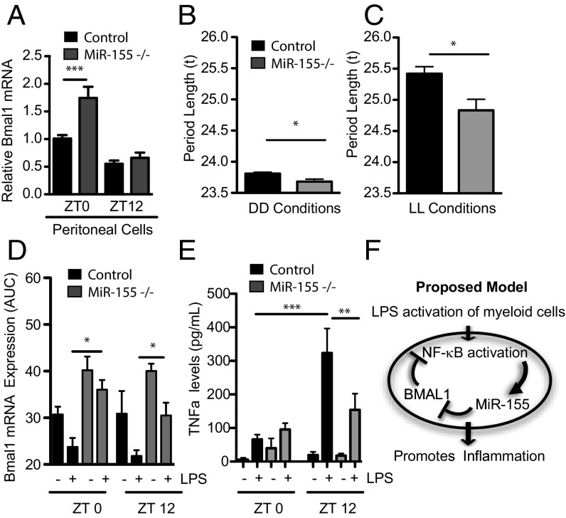
The response to an innate immune challenge is conditioned by the time of day, but the molecular basis for this remains unclear. In myeloid cells, there is a temporal regulation to induction by lipopolysaccharide (LPS) of the proinflammatory microRNA miR-155 that correlates inversely with levels of BMAL1. BMAL1 in the myeloid lineage inhibits activation of NF-κB and miR-155 induction and protects mice from LPS-induced sepsis. Bmal1 has two miR-155-binding sites in its 3'-UTR, and, in response to LPS, miR-155 binds to these two target sites, leading to suppression of Bmal1 mRNA and protein in mice and humans. miR-155 deletion perturbs circadian function, gives rise to a shorter circadian day, and ablates the circadian effect on cytokine responses to LPS. Thus, the molecular clock controls miR-155 induction that can repress BMAL1 directly. This leads to an innate immune response that is variably responsive to challenges across the circadian day.

Keywords: Bmal1; circadian clock; inflammation; miR-155; sepsis.


Conflict of interest statement

The authors declare no conflict of interest.

Figures

Fig. 1.
Fig. 1.
The magnitude of the circadian response to sepsis and inflammation correlates with induction of the microRNA miR-155. (A) Bmal1+/+Lys-MCre (n = 12–18) or (B) Bmal1−/−Lys-MCre male mice (n = 12–18) were injected intraperitoneally with LPS (25 mg/kg) at ZT0 (blue line) or ZT12 (red line) and monitored for survival over 7 d. Wild-type peritoneal cells harvested at ZT0 and ZT12 and treated immediately ex vivo with LPS (100 ng/mL) for indicated times and analyzed for expression of (C) mature miR-155 and (D) MiR-155HG (n = 3–4). Peritoneal cells harvested at ZT0 from Bmal1+/+Lys-MCre and Bmal1−/−Lys-MCre mice and treated immediately ex vivo with LPS (100 ng/mL) for the indicated time and analyzed for expression of (E) miR-155 (n = 3–4). (F) Serum-shocked BMDMs from Bmal1+/+Lys-MCre and Bmal1−/−Lys-MCre treated with LPS (100 ng/mL) for the indicated time and analyzed for expression of miR-155 (n = 3). (G) Peritoneal cells harvested at ZT0 from Bmal1+/+Lys-MCre and Bmal1−/−Lys-MCre mice, treated immediately ex vivo with LPS (1 ng/mL) for indicated times, and analyzed by immunoblot for levels of phosphorylated p65 at Serine 536 and total p65. Blot is representative of n = 6. Values provided are relative band intensity of phospho/total 65. Peritoneal cells as in E were analyzed for protein levels of (H) IL10 and (I) TNFα. *P ≤ 0.05 and ***P ≤ 0.001.
Fig. 2.
Fig. 2.
Activation of macrophages represses the core clock gene Bmal1, coincident with up-regulation of MiR-155. (A) Wild-type peritoneal cells harvested at ZT0 and ZT12 and treated immediately ex vivo with LPS (100 ng/mL) for 4 h and analyzed for Bmal1 (n = 3). (B) BMDMs were exposed to LPS (100 ng/mL) for the indicated times and analyzed for Bmal1 (n = 4–5). (C) BMAL1 protein by immunoblot (representative of n = 3). Values provided are relative band intensity corrected for β-actin. (D) BMDMs were treated with LPS (100 ng/mL), Poly-I:C (PIC, 100 µg/mL), Pam3CSK4 (100 ng/mL), R848 (1 µg/mL). and CpG (1 µg/mL) for 4 h and analyzed for Bmal1 (n = 3). RAW-264 cells exposed to E. coli or Salmonella for 24 h and analyzed for expression of (E) miR-155 and (F) Bmal1 (n = 3). (G) BMDMs from wild-type and Rev-Erbα −/− mice were treated with LPS (100 ng/mL) for 4 h and analyzed for Bmal1 (n = 3). BMDMs harvested from control and Il-10−/− mice and analyzed for expression of (H) miR-155 and (I) Bmal1 (n = 8–10). *P ≤ 0.05 and **P ≤ 0.01.
Fig. 3.
Fig. 3.
Bmal1 is repressed by the microRNA MiR-155 under basal and LPS conditions. (A) Schematic of Bmal1 3′ UTR illustrating position of the two miR-155–binding sites identified with the software TargetScan. iBMDMs transfected with a miR-155 mimic and analyzed for expression of (B) Bmal1 mRNA and (C) protein (n = 3). NC, negative control for mimic; LF, Lipofectamine; MiR-155O/E, overexpression of MiR-155 mimic. iBMDMs transfected with either a negative control for antagomir (NC) or an antagomir (Ant) to miR-155, treated with LPS (100 ng/mL), and analyzed for expression of (D) Bmal1 and (E) TNFα levels by ELISA (n = 3). WT or miR-155−/− BMDMs treated with LPS (100 ng/mL) for the indicated time and analyzed for expression of (F) Bmal1 and (G) protein levels by immunoblot. (H) Densitometry values of immunoblots from G and Fig. S4 (n = 3–4), and (I) Tnfα mRNA (n = 3). BMDMs transfected with a scrambled control morpholino (Scr), MiR-155 antagomir (Ant.) morpholino against the MiR-155 site in Bmal1 at position 40 (site 1), and morpholino against the MiR-155 site in Bmal1 at position 235 (site 2) were treated with LPS (100 ng/mL) for 24 h and analyzed for expression of (J) Bmal1 and (K) MiR-155. (L) Luciferase reporter activity from Bmal1 3′ UTR construct with LPS induction (100 ng/mL) or overexpression of MiR-155 mimic with wild-type luciferase construct or double (Mut. Site 1 + 2) mutations of miR-155–binding sites (n = 3). C, control; NC, negative control for mimic; 155, overexpression of miR-155 mimic. *P ≤ 0.05, **P ≤ 0.01, and ***P ≤ 0.001.
Fig. 4.
Fig. 4.
MiR-155 targets BMAL1 in humans and suppresses NF-κB activity. Human macrophages from four donors stimulated ex vivo with LPS (100 ng/mL) for the indicated times and analyzed for expression of (A) BMAL1 mRNA, (B) BMAL1 protein, and (C) miR-155. (D) hPBMCs were transfected with an antagomir to miR-155 or scrambled control and then treated with LPS for indicated times and immunobloted for BMAL1 and β-actin as control. Values provided are relative band intensity of BMAL1/β-Actin. (E) hPBMCs were transfected with an antagomir to miR-155 or scrambled control and then treated with LPS for indicated times and immunoblotted for phosphorylated p65 at Serine 536 and total p65 as control. Values provided are relative band intensity of phospho-p65/total p65. *P ≤ 0.05.
Fig. 5.
Fig. 5.
Altered clock function and clock gated cytokine responses in mice lacking MiR-155. (A) Peritoneal cells harvested from WT and miR-155−/− mice at ZT0 or ZT12 were immediately lysed and analyzed for Bmal1 (n = 3–4). Period length of control and miR-155–deficient mice in (B) constant dark conditions (DD) and (C) constant light conditions (LL) (n = 9–14). (D) Peritoneal macrophages harvested from WT and miR-155 mice at ZT0 and at ZT12 and treated immediately ex vivo with LPS (100 ng/mL) for 4, 8, and 24 h and analyzed for expression of Bmal1 by area under a curve (AUC). (E) Peritoneal macrophages were harvested from WT and miR-155−/− mice at ZT0 and at ZT12 and treated immediately ex vivo with LPS (100 ng/mL) for 24 h, and supernatants were analyzed by ELISA for TNFα (n = 4). (F) Schematic model to depict the circadian effect on LPS activation via the circadian control of miR-155 on Bmal1 in myeloid cells. *P ≤ 0.05, **P ≤ 0.01, and ***P ≤ 0.001.

References

    1. Yang G, et al. Knitting up the raveled sleave of care. Sci Transl Med. 2013;5(212):212rv213. - PubMed
    1. Curtis AM, Bellet MM, Sassone-Corsi P, O’Neill LA. Circadian clock proteins and immunity. Immunity. 2014;40(2):178–186. - PubMed
    1. Halberg F, Johnson EA, Brown BW, Bittner JJ. Susceptibility rhythm to E. coli endotoxin and bioassay. Proc Soc Exp Biol Med. 1960;103:142–144. - PubMed
    1. Nguyen KD, et al. Circadian gene Bmal1 regulates diurnal oscillations of Ly6C(hi) inflammatory monocytes. Science. 2013;341(6153):1483–1488. - PMC - PubMed
    1. Shackelford PG, Feigin RD. Periodicity of susceptibility to pneumococcal infection: Influence of light and adrenocortical secretions. Science. 1973;182(4109):285–287. - PubMed

Publication types

LinkOut - more resources