. 2015 Jul 15;26(14):2579-95.
doi: 10.1091/mbc.E15-03-0136. Epub 2015 May 20.

P-body assembly requires DDX6 repression complexes rather than decay or Ataxin2/2L complexes

Affiliations

P-body assembly requires DDX6 repression complexes rather than decay or Ataxin2/2L complexes

Jessica Ayache et al. Mol Biol Cell. .

Abstract

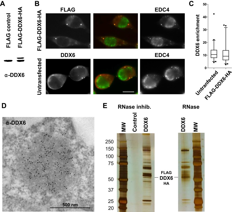
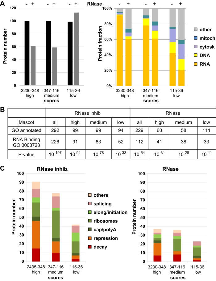
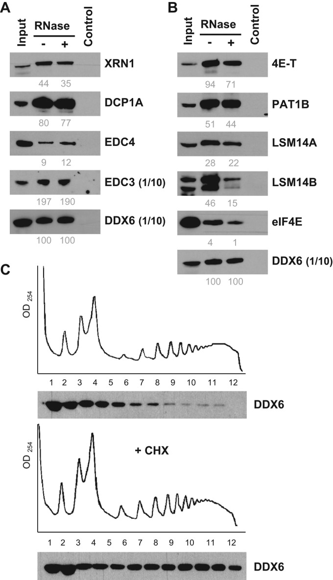
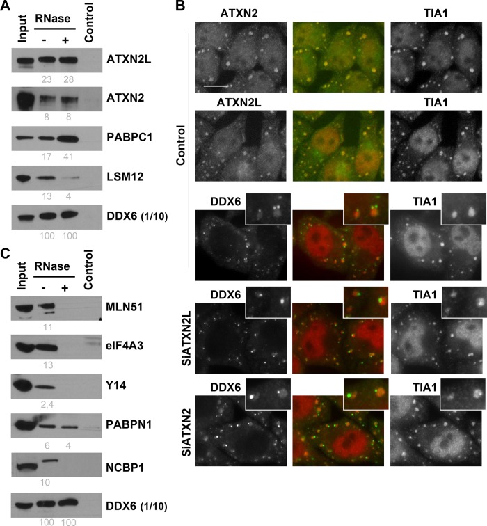
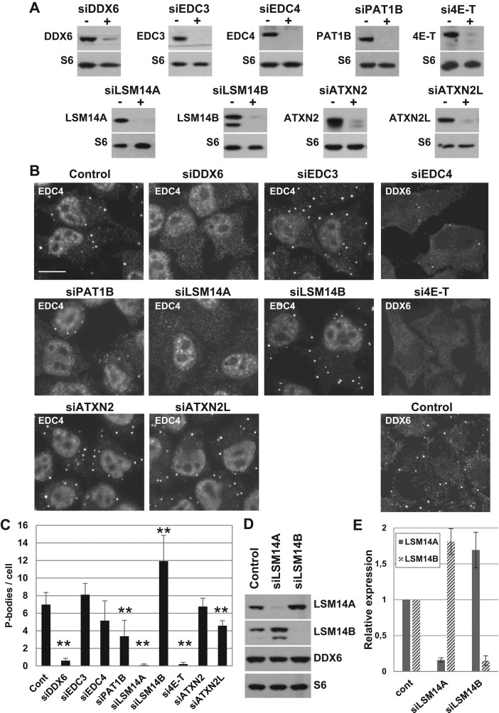
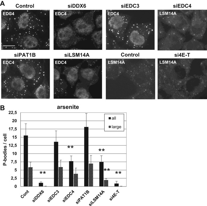
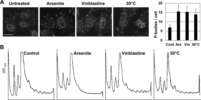
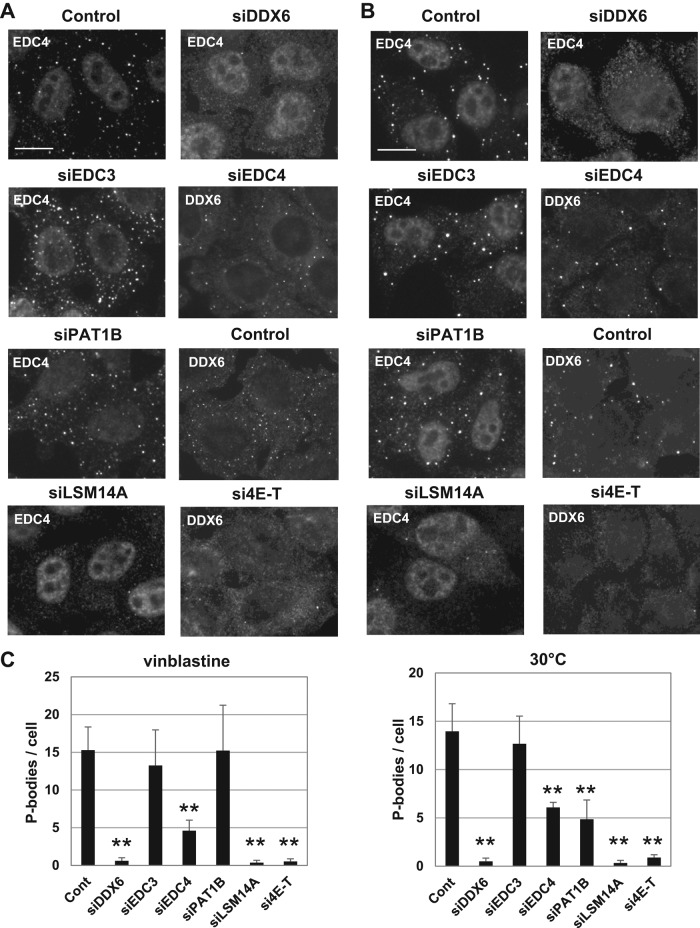
P-bodies are cytoplasmic ribonucleoprotein granules involved in posttranscriptional regulation. DDX6 is a key component of their assembly in human cells. This DEAD-box RNA helicase is known to be associated with various complexes, including the decapping complex, the CPEB repression complex, RISC, and the CCR4/NOT complex. To understand which DDX6 complexes are required for P-body assembly, we analyzed the DDX6 interactome using the tandem-affinity purification methodology coupled to mass spectrometry. Three complexes were prominent: the decapping complex, a CPEB-like complex, and an Ataxin2/Ataxin2L complex. The exon junction complex was also found, suggesting DDX6 binding to newly exported mRNAs. Finally, some DDX6 was associated with polysomes, as previously reported in yeast. Despite its high enrichment in P-bodies, most DDX6 is localized out of P-bodies. Of the three complexes, only the decapping and CPEB-like complexes were recruited into P-bodies. Investigation of P-body assembly in various conditions allowed us to distinguish required proteins from those that are dispensable or participate only in specific conditions. Three proteins were required in all tested conditions: DDX6, 4E-T, and LSM14A. These results reveal the variety of pathways of P-body assembly, which all nevertheless share three key factors connecting P-body assembly to repression.


Figures

FIGURE 1:
FIGURE 1:
Purification of DDX6 complexes by TAP-tag. (A) HEK293 cells were transfected with a FLAG-DDX6-HA or an empty plasmid as a control. After 48 h, proteins were analyzed by Western blotting with anti-DDX6 antibodies. (B) In parallel, transfected cells were stained with anti-FLAG (red) and anti-EDC4 (green) antibodies to detect the exogenous DDX6 protein and P-bodies, respectively. For comparison, untransfected cells were stained with anti-DDX6 and anti-EDC4 antibodies. Owing to the round shape of the cells, the maximal projection of a z-series of images is shown to better visualize P-bodies. Scale bar: 10 μm. (C) DDX6 enrichment in P-bodies with respect to the surrounding cytoplasm (n = 94 and 90 for transfected and untransfected samples, respectively) was quantified from single-plane images. (D) Endogenous DDX6 was detected in untransfected HEK293 cells by immunoelectron microscopy, as previously described (Souquere et al., 2009). One P-body highly enriched in DDX6 is shown. (E) Cells transfected with a FLAG-DDX6-HA or an empty plasmid were lysed in the presence of RNase inhibitor or RNase, and DDX6 complexes were purified by TAP-tag. Proteins were migrated on a denaturing gel along with a molecular weight marker (MW) and silver stained.
FIGURE 2:
FIGURE 2:
Functional description of the proteins identified by mass spectrometry. (A) Proteins purified from lysates treated with RNase inhibitor (− RNase) or RNase (+ RNase) were ranked by Mascot score and arbitrarily divided into three groups of high, medium, and low scores, as shown in the left panel. Their distribution into functional categories is based on the literature, and shown in the right panel. (B) RNA-binding proteins were searched using gene ontology. The table presents for each group (all, high, medium, low Mascot scores), the number of proteins annotated in the GO database (GO annotated), the number of RNA-binding annotated proteins (GO 0003723), and the p value associated with their enrichment. (C) Proteins categorized in RNA metabolism in A were subcategorized into indicated functions based on the literature.
FIGURE 3:
FIGURE 3:
Characterization of decay and repression complexes in untransfected cells. (A and B) HEK293 cytoplasmic lysates were immunoprecipitated with anti-DDX6 antibodies and analyzed by Western blotting for components of (A) decapping and (B) repression complexes, as indicated. For DDX6 and EDC3, only one-tenth of the immunoprecipitate was loaded on the gel to avoid saturation. For facilitation of comparison, the immunoprecipitation efficiency was estimated from scanned images, as described in Materials and Methods, and indicated below as percent values. (C) Cytoplasmic lysates from HEK293 incubated or not with cycloheximide (+ CHX) were separated by centrifugation on sucrose gradients and analyzed by optical densitometry (top panels). Proteins from collected fractions were analyzed by Western blotting with anti-DDX6 antibodies (bottom panels).
FIGURE 4:
FIGURE 4:
Characterization of the ATXN2/2L complex and EJC in untransfected cells. (A) Components of the ATXN2/2L complex were analyzed as in Figure 3, A and B. (B) HeLa cells were transfected with indicated siRNAs. After 48 h, cells were stressed with arsenite for 30 min. Localization of ATXN2, ATXN2L, and DDX6 in stress granules was analyzed by immunofluorescence, using TIA1 as a stress granule marker. Scale bar: 10 μm. (C) Components of the EJC were analyzed as in A.
FIGURE 5:
FIGURE 5:
Role of P-body proteins in the maintenance of P-bodies. (A–C) HeLa cells were transfected with indicated siRNAs. After 48 h, (A) proteins were analyzed by Western blotting with indicated antibodies, and (B) P-bodies were analyzed by immunofluorescence with anti-EDC4 or DDX6 antibodies, along with antibodies directed against the silenced protein to check the silencing at the individual cell level (unpublished data). Scale bar: 10 μm. (C) P-bodies were counted in three independent experiments, and their average number per cell was plotted. **, p < 0.005. (D and E) After transfection of siLSM14A and siLSM14B, proteins were analyzed by Western blotting using indicated antibodies (D). Signals were quantified in three independent experiments and plotted (E).
FIGURE 6:
FIGURE 6:
Role of P-body proteins in P-body assembly after arsenite treatment. HeLa cells were transfected with indicated siRNAs and analyzed as in Figure 5, except that cells were treated with arsenite for 30 min before fixation. P-bodies were immunostained (A) and counted (B) as in Figure 5 (all), except that LSM14A antibodies were used in place of DDX6, as this accumulated in both stress granules and P-bodies. A second quantitation restricted to P-bodies larger than 450 nm was performed to show the characteristic defect observed after silencing LSM14A (large). Scale bar: 10 μm. The corresponding Western blot analysis is presented in Supplemental Figure 2D.
FIGURE 7:
FIGURE 7:
P-body induction after treatment with arsenite, vinblastine, and mild cold shock. HeLa cells were treated with arsenite for 30 min, vinblastine for 1 h, or cultured at 30°C for 2 h before lysis or fixation. (A) P-bodies were detected and counted as in Figure 5. Scale bar: 10 μm. (B) Cytoplasmic lysates were separated on sucrose gradients and analyzed by optic densitometry, as in Figure 3C.
FIGURE 8:
FIGURE 8:
Role of P-body proteins in P-body assembly after vinblastine treatment and mild cold shock. HeLa cells were transfected with indicated siRNAs and analyzed, as in Figure 5, except that cells were treated with vinblastine (A) or cultured at 30°C (B) before fixation. P-bodies were counted as in Figure 5 (C). Scale bar: 10 μm. The corresponding Western blot analysis is presented in Supplemental Figure 2D.

References

    1. Aizer A, Brody Y, Ler LW, Sonenberg N, Singer RH, Shav-Tal Y. The dynamics of mammalian P body transport, assembly, and disassembly in vivo. Mol Biol Cell. 2008;19:4154–4166. - PMC - PubMed
    1. Andrei MA, Ingelfinger D, Heintzmann R, Achsel T, Rivera-Pomar R, Lührmann R. A role for eIF4E and eIF4E-transporter in targeting mRNPs to mammalian processing bodies. RNA. 2005;11:717–727. - PMC - PubMed
    1. Buchan JR. mRNP granules: assembly, function, and connections with disease. RNA Biol. 2014:e29034. - PMC - PubMed
    1. Carbonaro M, O'Brate A, Giannakakou P. Microtubule disruption targets HIF-1α mRNA to cytoplasmic P-bodies for translational repression. J Cell Biol. 2011;192:83–99. - PMC - PubMed
    1. Chen Y, Boland A, Kuzuog˘lu-Öztürk D, Bawankar P, Loh B, Chang C-T, Weichenrieder O, Izaurralde E. A DDX6-CNOT1 complex and W-binding pockets in CNOT9 reveal direct links between miRNA target recognition and silencing. Mol Cell. 2014;54:737–750. - PubMed

Publication types

MeSH terms