. 2015 Jul 15;26(14):2673-84.
doi: 10.1091/mbc.E15-04-0224. Epub 2015 May 20.

Endosomal regulation of contact inhibition through the AMOT:YAP pathway

Affiliations

Endosomal regulation of contact inhibition through the AMOT:YAP pathway

Christopher M Cox et al. Mol Biol Cell. .

Abstract

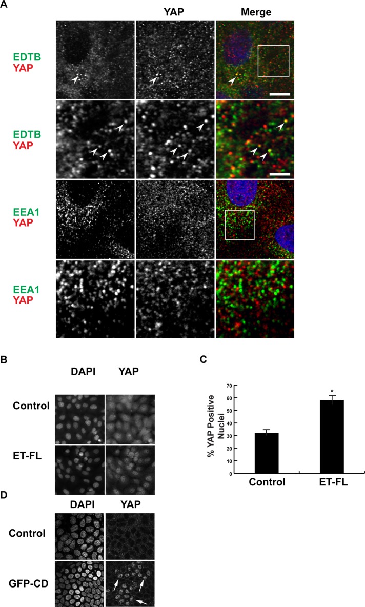
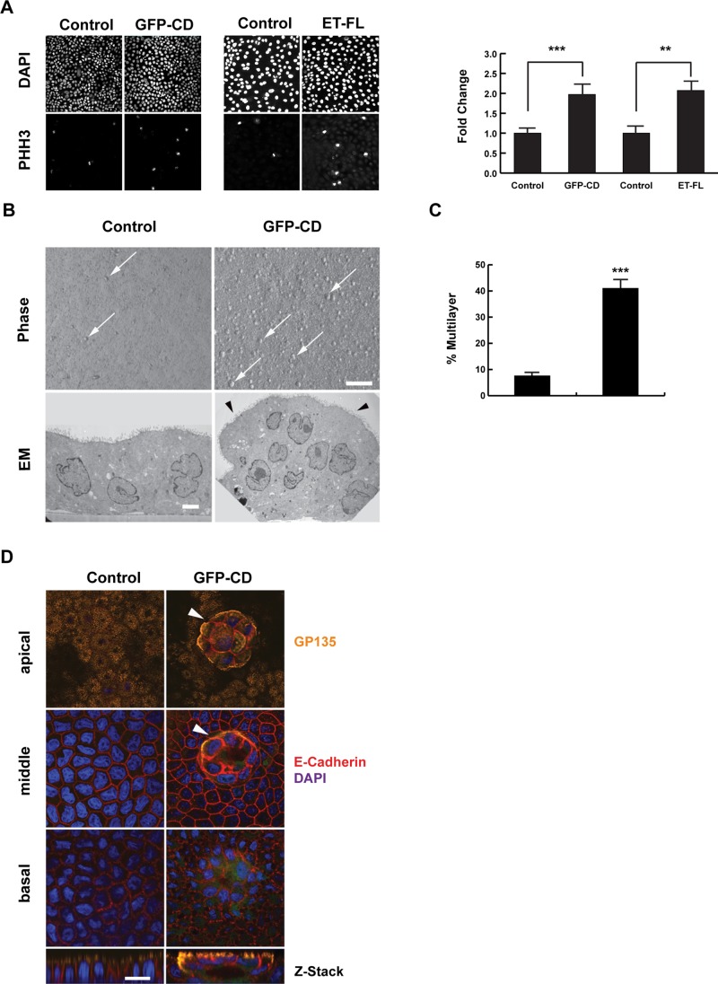
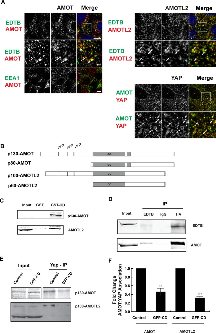
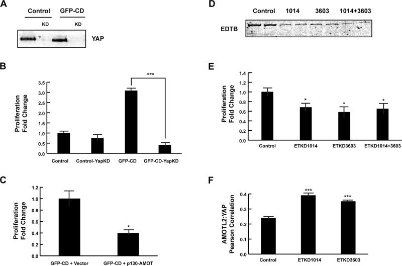
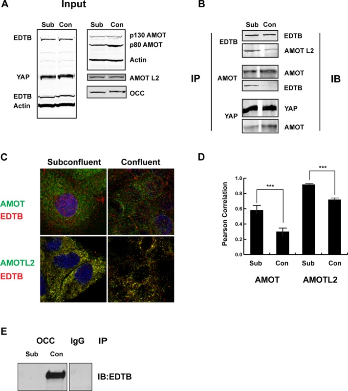
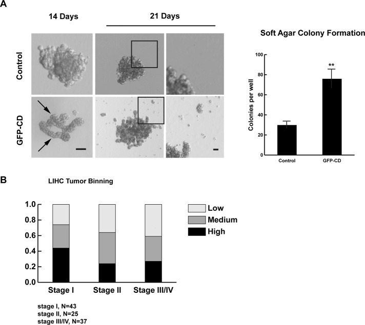
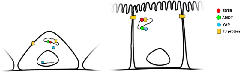
Contact-mediated inhibition of cell proliferation is an essential part of organ growth control; the transcription coactivator Yes-associated protein (YAP) plays a pivotal role in this process. In addition to phosphorylation-dependent regulation of YAP, the integral membrane protein angiomotin (AMOT) and AMOT family members control YAP through direct binding. Here we report that regulation of YAP activity occurs at the endosomal membrane through a dynamic interaction of AMOT with an endosomal integral membrane protein, endotubin (EDTB). EDTB interacts with both AMOT and occludin and preferentially associates with occludin in confluent cells but with AMOT family members in subconfluent cells. EDTB competes with YAP for binding to AMOT proteins in subconfluent cells. Overexpression of the cytoplasmic domain or full-length EDTB induces translocation of YAP to the nucleus, an overgrowth phenotype, and growth in soft agar. This increase in proliferation is dependent upon YAP activity and is complemented by overexpression of p130-AMOT. Furthermore, overexpression of EDTB inhibits the AMOT:YAP interaction. EDTB and AMOT have a greater association in subconfluent cells compared with confluent cells, and this association is regulated at the endosomal membrane. These data provide a link between the trafficking of tight junction proteins through endosomes and contact-inhibition-regulated cell growth.


Figures

FIGURE 1:
FIGURE 1:
YAP and EDTB colocalize on endosomes. (A) MDCK cells grown on coverslips were labeled for endogenous EDTB, YAP, and EEA1. YAP colocalizes with EDTB (arrowheads) on intracellular puncta with a Pearson r of 0.31, whereas the Pearson r of YAP with EEA1 is 0.18. Scale bars, 10 μm (top), 2.5 μm (inset). (B–D) EDTB overexpression induces enrichment of YAP in the nucleus. MDCK cells expressing full-length EDTB (B, ET-FL) or control plasmid were grown on coverslips to confluence, and YAP localization was assessed using immunofluorescence. DAPI staining was used to determine total cell number. Representative images. (C) Quantification of B. ImageJ was used for quantification of nuclear enrichment of YAP protein. An identical threshold was set for all images, and YAP-positive nuclei were counted using the analyze particles function of ImageJ. Error bars, SEM of percentage of cells with nuclear enrichment of YAP; *p < 0.05. (D) GFP-CD and control cells grown to confluence and labeled with YAP. Expression of GFP-CD results in increased nuclear YAP (arrows).
FIGURE 2:
FIGURE 2:
EDTB overexpression results in loss of growth control. (A) Representative cells expressing ET-FL or GFP-CD labeled with PHH3. Proliferation is increased with overexpression of GFP-CD or ET-FL. Error bars, SEM of percentage of cells that are PHH3 positive; ***p < 0.005, **p < 0.01. (B) GFP-CD cells form multiple small, multilayered foci (arrows) when grown on Transwell filters, whereas control cells remain as a monolayer with limited number of foci. Transmission electron microscopy (TEM) of control and GFP-CD cells grown on filters shows that control cells grow as a monolayer but GFP-CD cells form multicellular foci. Foci of GFP-CD–expressing cells maintain epithelial characteristics, such as apical microvilli (arrowheads). Scale bars, 100 μm (phase), 1 μm (TEM). (C) Quantification of the multilayer area shows an increase in foci formation in cells expressing GFP-CD. Error bars, SEM; ***p < 0.005. (D) Immunofluorescence labeling of apical (gp135, podocalyxin) and basolateral (E-cadherin) markers shows that the polarized distribution of these proteins is maintained with expression of GFP-CD. The foci (arrowheads) also maintain this polarized expression pattern. Scale bar, 20 μm.
FIGURE 3:
FIGURE 3:
EDTB, AMOT, and YAP all localize to endosomes, and EDTB regulates AMOT interaction with YAP. (A) Localization of endogenous AMOT, AMOTL2, YAP, EDTB, and EEA1 in MDCK cells grown on coverslips. EDTB colocalizes with AMOT (Pearson's r = 0.65) and AMOTL2 (Pearson's r = 0.79) on intracellular puncta (arrows), but there is limited colocalization with EEA1 (Pearson's r for AMOT/EEA1 is 0.18). AMOT and YAP also colocalize on intracellular puncta (Pearson's r = 0.56). Scale bars, 5 μm (top), 1 μm (insets). (B) AMOT and AMOTL2 domain structure. p80-AMOT and p60-AMOTL2 isoforms result from an internal start site. The amino-terminal domains of p130-AMOT and p100-AMOTL2 interact with YAP through the PPxY motif. (C) MDCK lysates collected at 90% confluence were used to assess the physical interaction of EDTB and AMOT/AMOTL2. Pull-down using the cytoplasmic domain of EDTB fused to GST (GST-CD) analyzed by Western blot shows interaction with AMOT and AMOTL2 relative to the GST-only control. AMOT input is sometimes not detected, indicating concentration by pull down. (D) Coimmunoprecipitation of ET-FL and HA-p130-AMOT. HEK293 cells were transfected with ET-FL and HA-p130AMOT. Lysates were immunoprecipitated with EDTB or HA antibodies. Western blot analysis shows coimmunoprecipitation of EDTB and AMOT. (E) Competition of EDTB with YAP for binding to AMOT and AMOTL2. MDCK cells expressing GFP-CD or control plasmid were immunoprecipitated with antibody against YAP and analyzed by Western blot for AMOT or AMOTL2. In the presence of GFP-CD, there is a significant decrease in the amount of AMOT and AMOTL2 associated with YAP compared with control. (F) Quantification of E. Error bars, SEM from three independent experiments. **p < 0.01, ***p < 0.005.
FIGURE 4:
FIGURE 4:
YAP activity is required for GFP-CD–induced increase in proliferation. (A) MDCK cells expressing GFP or GFP-CD were infected with YAP shRNA lentivirus. Lysates were analyzed by Western blot for expression of YAP. YAP protein levels are decreased >90% in both the control and GFP-CD–expressing cells. (B) Changes in proliferation were determined by PHH3 labeling. Proliferation of GFP-CD cells is significantly decreased with YAP knockdown. Error bars, SEM; ***p < 0.005. YAP knockdown in control cells also resulted in decreased proliferation (0.74-fold of that of parent lines). (C) MDCK cells expressing GFP-CD were transfected with p130AMOT. Overexpression of p130-AMOT complements the GFP-CD increased-proliferation phenotype. Error bars, SEM; *p < 0.05. (D) Endogenous EDTB was knocked down in MDCK cells using two independent shRNAs, individually or together. Lysates were analyzed by Western blot to verify the reduction in EDTB expression. (E) Proliferation is decreased when EDTB expression is reduced. Error bars, SEM; *p < 0.05. (F) Pearson r of AMOTL2:YAP colocalization in EDTB knockdown cells. Knockdown of endotubin results in a significant increase in AMOTL2:YAP colocalization. Four to six images were quantified for each condition. Error bars, SEM; ***p < 0.005.
FIGURE 5:
FIGURE 5:
Interaction between EDTB and AMOT is regulated by cell density. (A) The expression levels of EDTB, AMOT, AMOTL2, YAP, and occludin in subconfluent and confluent MDCK cultures were analyzed by Western blot analysis. (B) Top, immunoprecipitation of MDCK subconfluent and confluent cultures with EDTB was analyzed by Western blot for AMOTL2. Middle, immunoprecipitation of MDCK subconfluent and confluent cultures with AMOT was analyzed by Western blot for EDTB. Bottom, immunoprecipitation of MDCK subconfluent and confluent cultures with YAP was analyzed by Western blot for AMOT. (C) MDCK cells plated on coverslips were immunolabeled for EDTB and AMOT or AMOTL2. (D) The amount of colocalization between AMOT/EDTB and AMOTL2/EDTB was determined by calculating the Pearson r. The colocalization of AMOT/EDTB and AMOTL2/EDTB is significantly reduced in confluent cultures. Average endosome number per image is 475, and eight images were quantified per condition. Error bars, SEM; ***p < 0.005. (E) Immunoprecipitation of MDCK subconfluent and confluent cultures with occludin was analyzed by Western blot for EDTB. The image is spliced from the same gel.
FIGURE 6:
FIGURE 6:
Increased expression of EDTB results in growth in soft agar and is associated with early-stage LIHC samples. (A) MDCK cells expressing GFP (control) or GFP-CD were grown in soft agar for 3 wk. GFP-CD cells form long, branching structures (arrows) that produce satellite colonies (arrowheads) after 3 wk in culture. Scale bars, 100 μm (left), 20 μm (right). Right, quantification of colony formation after 14 d in cells expressing GFP-CD. Error bars, SEM; **p < 0.01. (B) Using the Cancer Genome Database, we collected the rsem expression values for EDTB in 105 liver hepatocellular carcinomas. The tumors were binned into three equal groups based on EDTB expression levels. Separating these groups by tumor stage shows that 44% of stage I tumors express EDTB at high levels.
FIGURE 7:
FIGURE 7:
Model for endosomal control of contact inhibition through YAP:AMOT. In subconfluent cells, AMOT associates with EDTB on endosomes, allowing YAP to localize to the nucleus. When cells reach confluence, tight junction proteins cycling through endosomes associate with EDTB, allowing AMOT to sequester YAP at the endosomal membrane.

References

    1. Alkasalias T, Flaberg E, Kashuba V, Alexeyenko A, Pavlova T, Savchenko A, Szekely L, Klein G, Guven H. Inhibition of tumor cell proliferation and motility by fibroblasts is both contact and soluble factor dependent. Proc Natl Acad Sci USA. 2014;111:17188–17193. - PMC - PubMed
    1. Archibald A, Mihai C, Macara IG, McCaffrey L. Oncogenic suppression of apoptosis uncovers a Rac1/JNK proliferation pathway activated by loss of Par3. Oncogene. 2014:doi:10.1038/onc.2014.242. - PMC - PubMed
    1. Bai H, Gayyed MF, Lam-Himlin DM, Klein AP, Nayar SK, Xu Y, Khan M, Argani P, Pan D, Anders RA. Expression of Yes-associated protein modulates Survivin expression in primary liver malignancies. Hum Pathol. 2012;43:1376–1385. - PMC - PubMed
    1. Basu S, Totty NF, Irwin MS, Sudol M, Downward J. Akt phosphorylates the Yes-associated protein, YAP, to induce interaction with 14-3-3 and attenuation of p73-mediated apoptosis. Mol Cell. 2003;11:11–23. - PubMed
    1. Boggiano JC, Fehon RG. Growth control by committee: intercellular junctions, cell polarity, and the cytoskeleton regulate Hippo signaling. Dev Cell. 2012;22:695–702. - PMC - PubMed

Publication types

MeSH terms

Substances

LinkOut - more resources