. 2015 Jul 15;26(14):2609-19.
doi: 10.1091/mbc.E14-07-1213. Epub 2015 May 20.

Cdc6 degradation requires phosphodegron created by GSK-3 and Cdk1 for SCFCdc4 recognition in Saccharomyces cerevisiae

Affiliations

Cdc6 degradation requires phosphodegron created by GSK-3 and Cdk1 for SCFCdc4 recognition in Saccharomyces cerevisiae

Amr Al-Zain et al. Mol Biol Cell. .

Abstract

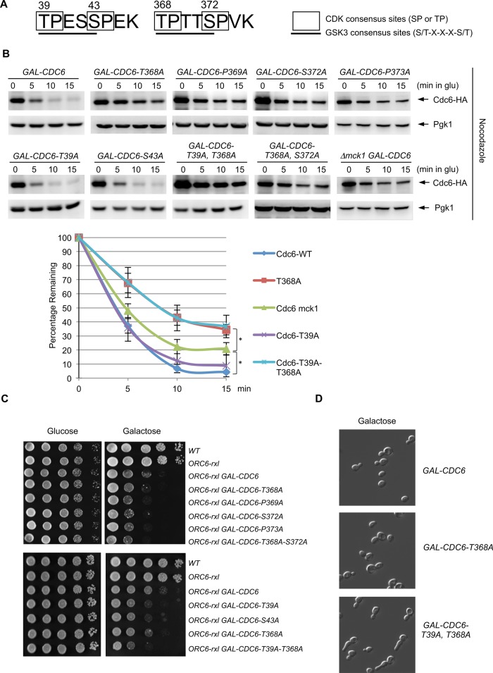
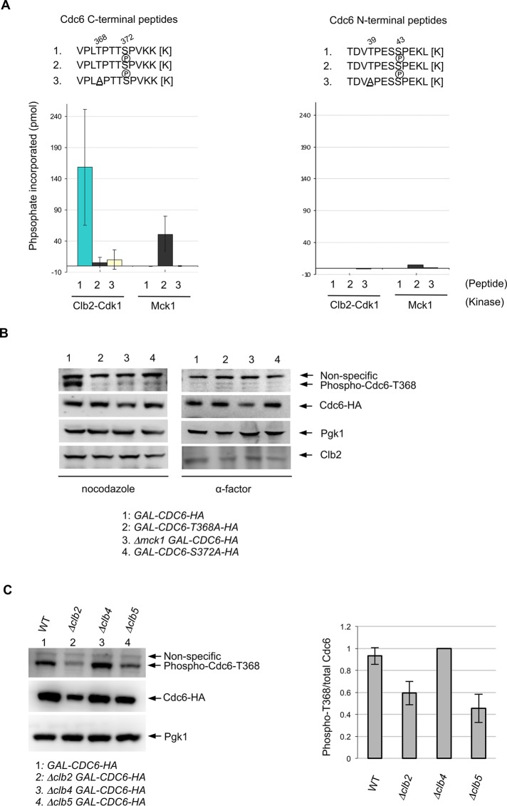
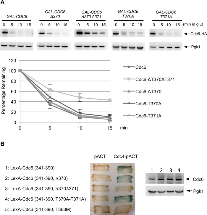
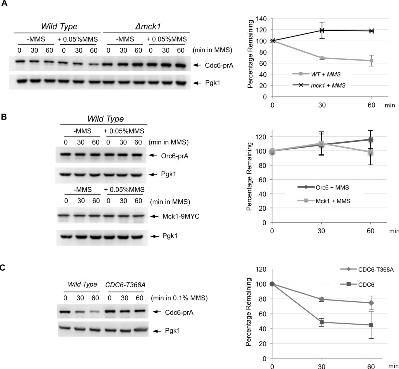
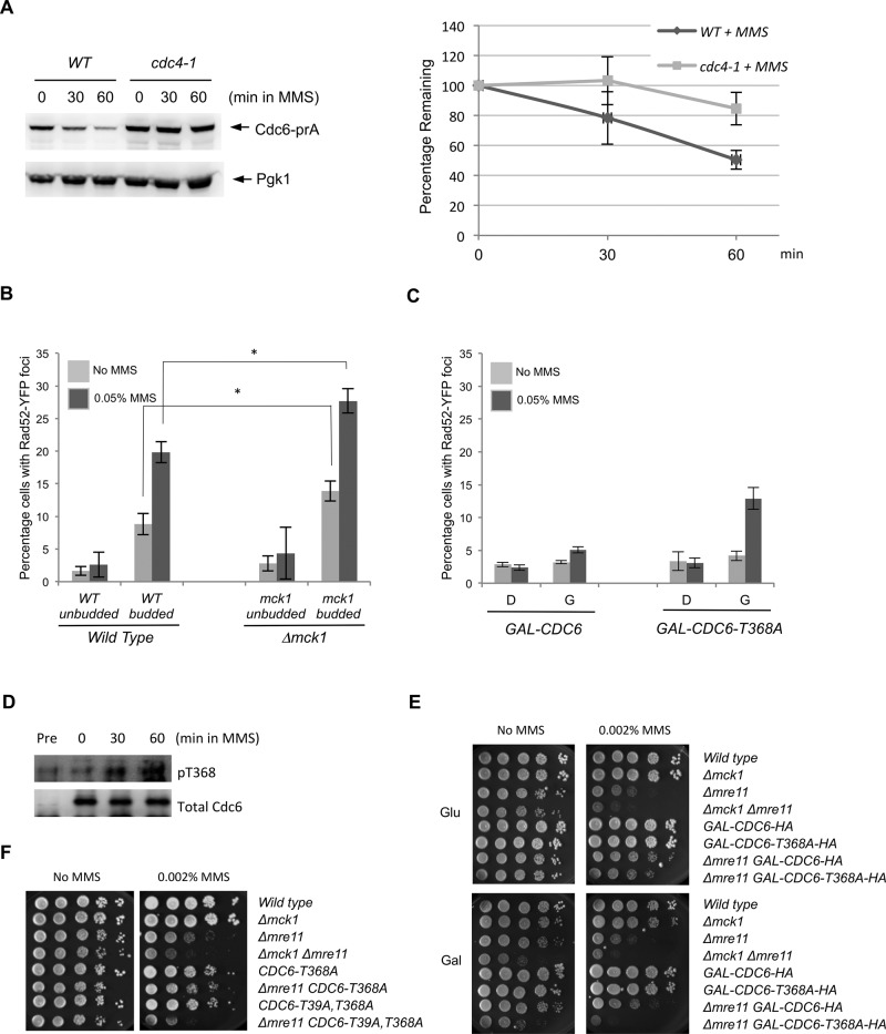
To ensure genome integrity, DNA replication takes place only once per cell cycle and is tightly controlled by cyclin-dependent kinase (Cdk1). Cdc6p is part of the prereplicative complex, which is essential for DNA replication. Cdc6 is phosphorylated by cyclin-Cdk1 to promote its degradation after origin firing to prevent DNA rereplication. We previously showed that a yeast GSK-3 homologue, Mck1 kinase, promotes Cdc6 degradation in a SCF(Cdc4)-dependent manner, therefore preventing rereplication. Here we present evidence that Mck1 directly phosphorylates a GSK-3 consensus site in the C-terminus of Cdc6. The Mck1-dependent Cdc6 phosphorylation required priming by cyclin/Cdk1 at an adjacent CDK consensus site. The sequential phosphorylation by Mck1 and Clb2/Cdk1 generated a Cdc4 E3 ubiquitin ligase-binding motif to promote Cdc6 degradation during mitosis. We further revealed that Cdc6 degradation triggered by Mck1 kinase was enhanced upon DNA damage caused by the alkylating agent methyl methanesulfonate and that the resulting degradation was mediated through Cdc4. Thus, Mck1 kinase ensures proper DNA replication, prevents DNA damage, and maintains genome integrity by inhibiting Cdc6.


Figures

FIGURE 1:
FIGURE 1:
Analysis of GSK-3 consensus sites in Cdc6. (A) Cdc6 contains two GSK-3 consensus sites, which overlap with two CDK consensus sites. (B) GAL-CDC6-HA strains with various mutations (T368A, P369A, S372A, P373A, T39A, S43A, T39A-T368A, and T368A-S372A) were expressed with galactose-containing medium for 2 h and then blocked with nocodazole for 2 h. Cdc6 expression was then suppressed by adding glucose. Protein extracts were collected every 5 min and subjected to Western blot analysis to observe Cdc6-HA. Pgk1 was used as a loading control. GAL-CDC6-HA in mck1-deletion cells was examined using the same method. Western blotting images for WT, Δmck1, CDC6-T39A, CDC6-T368A, and CDC6-T39A,T368A were quantified. Percentage of Cdc6 protein remaining relative to time zero is shown. Results are the average of three independent experiment, and error bars indicate SD. *p < 0.05. (C) Strains with the indicated genotypes were serially diluted 10-fold, plated on yeast extract/peptone/dextrose or yeast extract/peptone/galactose plates, and incubated at 30°C for 2 d. (D) GAL-CDC6, GAL-CDC6-T368A, or GAL-CDC6-T39A-T368A was grown in raffinose-containing medium first. Cdc6p was expressed with galactose for 3 h.
FIGURE 2:
FIGURE 2:
Mck1 phosphorylates the T368 site in Cdc6 after priming by Clb2-Cdk1. (A) To measure Clb2-Cdk1 and Mck1 kinase activities on Cdc6 phosphopeptides, various synthetic peptides of Cdc6 (residues 36–47 or 365–376; shown above) were incubated with purified kinases from asynchronous yeast cultures and [γ-32P]ATP. For each kinase, phosphate incorporation was normalized to a control reaction without peptides. Values for the C-terminal peptides represent the average from three independent experiments. Error bars represent SD. (B) Indicated strains were grown in raffinose-containing medium first. Cdc6 expression was induced with galactose for 2 h. Cells were blocked with nocodazole or α-factor for 2 h. Western blotting was performed using anti–phosphoT368 of Cdc6, anti-HA, anti-Pgk1, and anti-Clb2 antibodies, respectively. (C) Indicated strains were grown in raffinose-containing medium first, and then galactose was added to induce Cdc6 expression for 2 h. Cells were blocked with nocodazole for 2 h. Western blotting was performed using anti–phosphoT368 of Cdc6, anti-HA, and anti-Pgk1 antibodies, respectively (left). Band intensity for the phospho-Cdc6-T368 was quantified and normalized to the total amount of Cdc6 (right). The ratio phospho-T368/total Cdc6 in Δclb4 was set as 1. Results are the average of three independent experiments; error bars indicate SD.
FIGURE 3:
FIGURE 3:
Cdc6 phosphodegron for Cdc4. (A) GAL-CDC6-HA strains with various mutations (Δ370Δ371, Δ370, T370A, or T371A) were incubated in raffinose-containing medium and transferred to galactose-containing medium for 2 h to induce Cdc6 expression, and then cells were blocked with nocodazole. Cdc6 expression was suppressed by adding glucose. Protein samples were collected every 5 min and subjected to Western blot analysis to observe Cdc6-HA. Pgk1 was used as a loading control. Western blotting images were quantified. Results represent the average of three experiments, with error bars indicating SD. (B) Yeast two-hybrid analysis was performed to determine the binding between Cdc4p and the Cdc6p C-terminus. Full-length CDC4 in the pACT plasmid, containing the GAL4 activation domain (GAD), was cotransformed into L40 yeast strains along with the various CDC6 mutants in the pBTM116 plasmid fused to LexA. Transformants were assayed for β-galactosidase activity, as visualized in blue. Proteins were extracted from each strain (1–4), and Cdc6 protein levels were examined by Western blotting.
FIGURE 4:
FIGURE 4:
DNA damage triggers Cdc6 degradation in Mck1-dependent manner. (A) CDC6-prA or Δmck1 CDC6-prA cells were incubated in yeast extract/peptone/dextrose (YEPD) medium to log phase, and then MMS was added (0.05% final). Protein extracts were made at 0-, 30-, or 60-min incubation and subjected to Western blotting to visualize endogenous Cdc6-prA. Pgk1 was used as a loading control. The same experiment was repeated three times to quantify Cdc6 protein levels. Average percentage of Cdc6p remaining is shown. Bars represent SD. (B) Protein degradation of Orc6-prA or Mck1-9MYC was examined by the same method using IgG or anti-MYC antibodies, respectively. (C) CDC6-prA or CDC6-T368A-prA cells were grown in YEPD to log phase. MMS at 0.1% concentration was added, and protein samples were collected after 0, 30, and 60 min. The protein extracts were subjected to Western blotting to visualize Cdc6-prA. The same experiment was performed three times. Cdc6 protein levels were quantified, and average percentage of Cdc6p remaining is shown. Bars represent SD.
FIGURE 5:
FIGURE 5:
Cdc6 degradation upon DNA damage is mediated through the ubiquitin pathway. (A) CDC6-prA or cdc4-1 CDC6-prA cells under the endogenous promoter were grown to log phase at 26°C first and then the temperature was increased to 36°C for 1.5 h. Then MMS was added (0.1% final). Samples were collected after 0, 30, or 60 min. The same experiment was performed three times, and Cdc6 protein levels were quantified. Error bars represent SD. (B) RAD52-YFP or Δmck1 RAD52-YFP cells were grown to log phase in low-fluorescence medium. MMS was added (0.05% final) for 90 min. Rad52-YFP foci were visualized under a fluorescence microscope to assay for double-stranded DNA breaks before and after treatment with MMS. Percentage of cells containing Rad52 foci was determined in unbudded or budded cells. One hundred cells were counted for each sample. Percentage is average from three independent experiments, and error bars represent the SD. *p < 0.05. (C) GAL-CDC6 or GAL-CDC6-T368A cells were grown to log phase in raffinose-containing medium first, and then either glucose or galactose was added for 3 h of incubation. Finally, MMS was then added (0.05% final) for 1 h. Rad52-YFP foci formation was determined with or without MMS treatment. One hundred cells were counted for each sample. Percentage is average from three independent experiments, and error bars represent SD. (D) cdc4-1 GAL-CDC6-HA cells were incubated in raffinose-containing medium first. Galactose was then added for 1 h of incubation, followed by nocodazole for 1.5 h. The temperature was then raised to 36°C to inactivate Cdc4 function for 1.5 h. Finally, cells were treated with MMS (0.1% final) and sampled every 30 min. Western blotting was performed using anti-phosphoT368 of Cdc6 and anti-HA. Control experiment was performed in raffinose-containing medium, indicated as “pre.” (E, F) Cells with indicated genotypes were serially diluted 10-fold on yeast extract/peptone/dextrose or yeast extract/peptone/galactose plates containing 0.002% MMS. The plates were incubated at 30°C for 2 d.

Similar articles

Cited by

References

    1. Andoh T, Hirata Y, Kikuchi A. Yeast glycogen synthase kinase 3 is involved in protein degradation in cooperation with Bul1, Bul2, and Rsp5. Mol Cell Biol. 2000;20:6712–6720. - PMC - PubMed
    1. Araki H. Cyclin-dependent kinase-dependent initiation of chromosomal DNA replication. Curr Opin Cell Biol. 2010;22:766–771. - PubMed
    1. Archambault V, Ikui AE, Drapkin BJ, Cross FR. Disruption of mechanisms that prevent rereplication triggers a DNA damage response. Mol Cell Biol. 2005;25:6707–6721. - PMC - PubMed
    1. Borlado LR, Mendez J. CDC6: from DNA replication to cell cycle checkpoints and oncogenesis. Carcinogenesis. 2008;29:237–243. - PubMed
    1. Boronat S, Campbell JL. Mitotic Cdc6 stabilizes anaphase-promoting complex substrates by a partially Cdc28-independent mechanism, and this stabilization is suppressed by deletion of Cdc55. Mol Cell Biol. 2007;27:1158–1171. - PMC - PubMed

Publication types

MeSH terms