. 2015 May 20;35(20):7695-714.
doi: 10.1523/JNEUROSCI.4383-14.2015.

Covert shifts of spatial attention in the macaque monkey

Affiliations

Covert shifts of spatial attention in the macaque monkey

Natalie Caspari et al. J Neurosci. .

Abstract

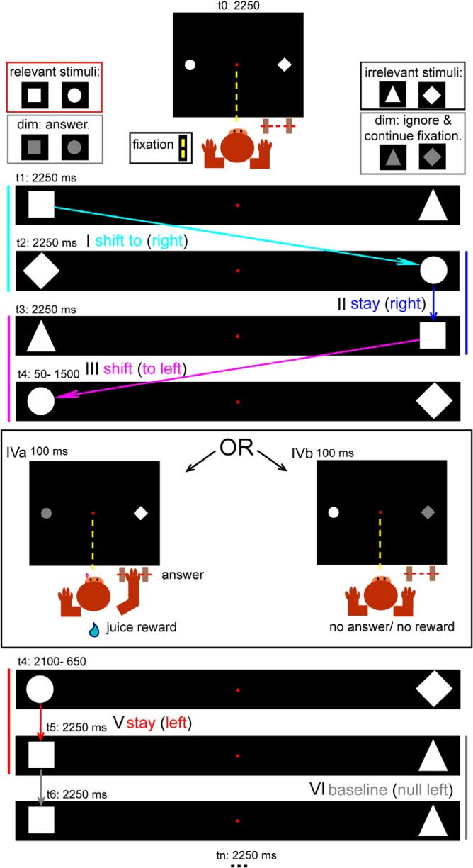
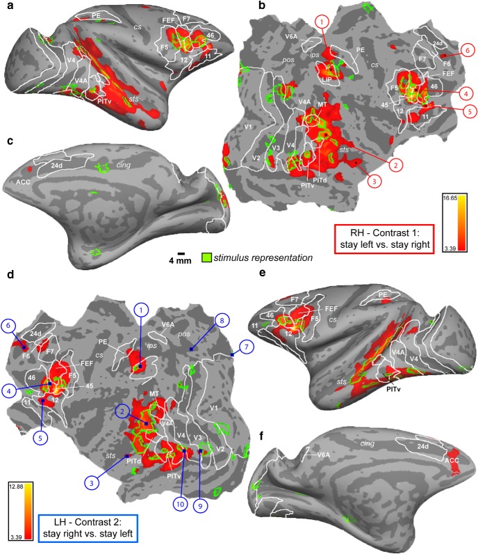
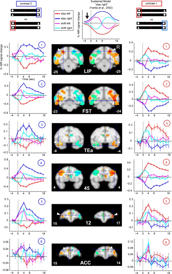
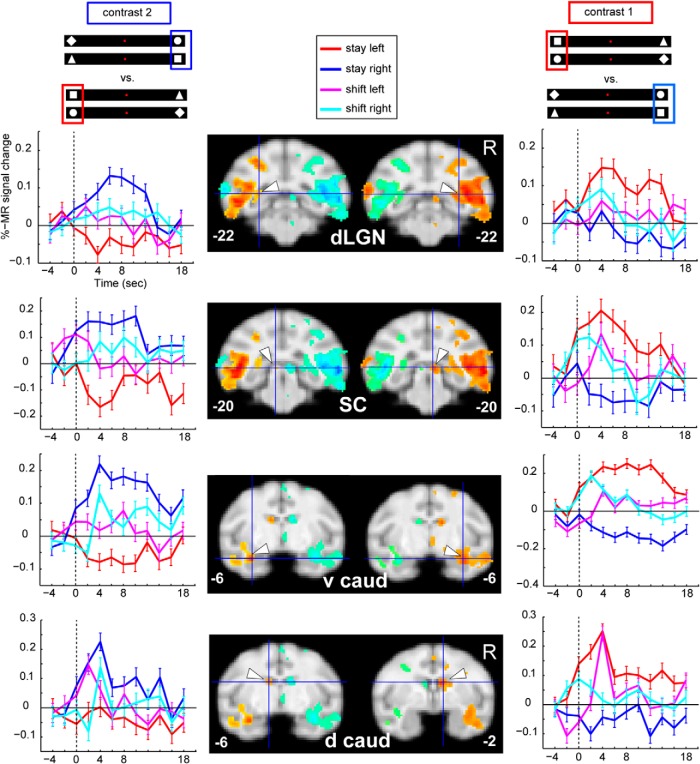
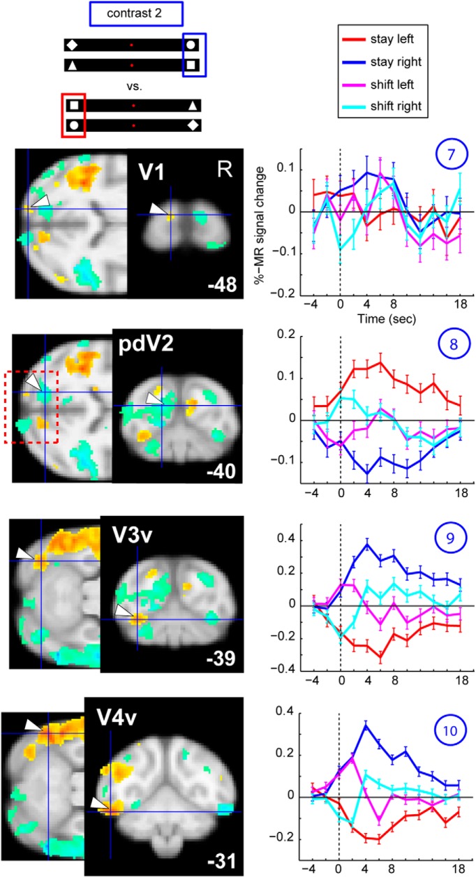
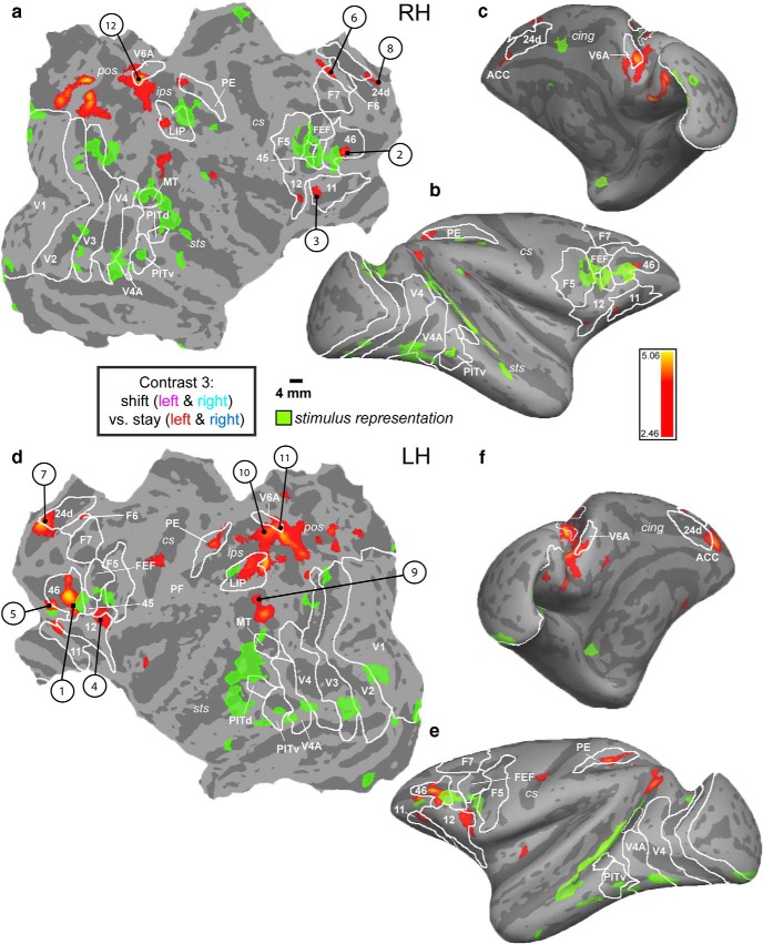
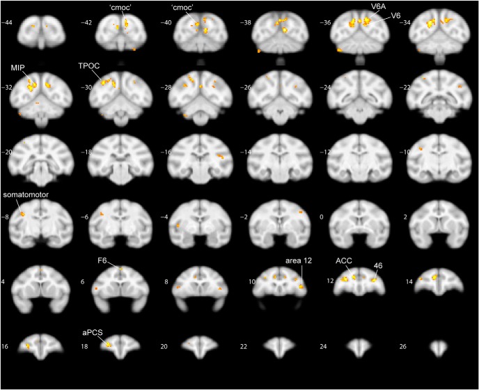
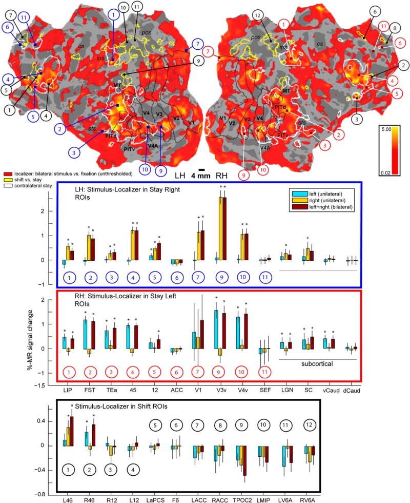
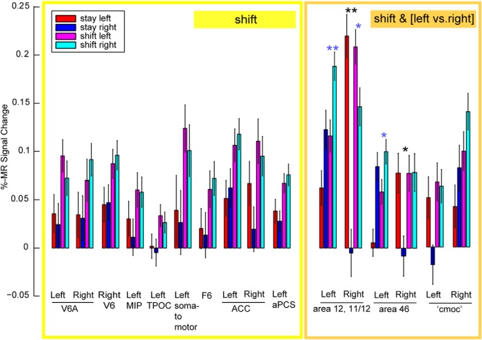
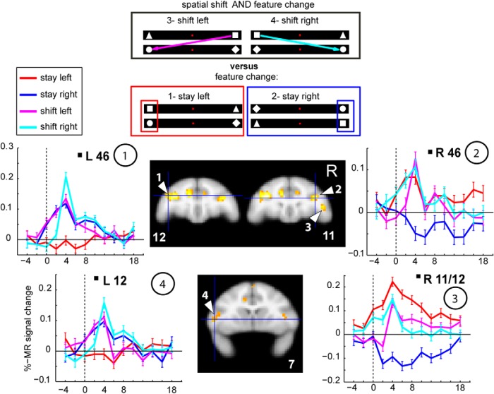
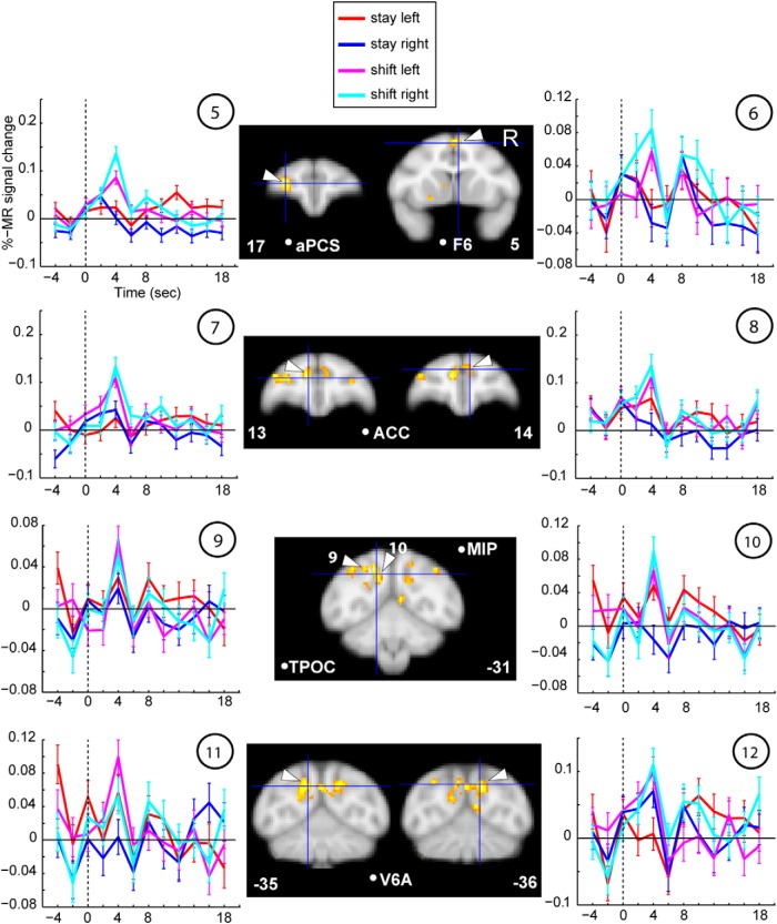
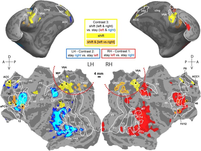
In the awake state, shifts of spatial attention alternate with periods of sustained attention at a fixed location or object. Human fMRI experiments revealed the critical role of the superior parietal lobule (SPL) in shifting spatial attention, a finding not predicted by human lesion studies and monkey electrophysiology. To investigate whether a potential homolog of the human SPL shifting region exists in monkeys (Macaca mulatta), we adopted an event-related fMRI paradigm that closely resembled a human experiment (Molenberghs et al., 2007). In this paradigm, a pair of relevant and irrelevant shapes was continuously present on the horizontal meridian. Subjects had to covertly detect a dimming of the relevant shape while ignoring the irrelevant dimmings. The events of interest consisted of the replacement of one stimulus pair by the next. During shift but not stay events, the relevant shape of the new pair appeared at the contralateral position relative to the previous one. Spatial shifting events activated parietal areas V6/V6A and medial intraparietal area, caudo-dorsal visual areas, the most posterior portion of the superior temporal sulcus, and several smaller frontal areas. These areas were not activated during passive stimulation with the same sensory stimuli. During stay events, strong direction-sensitive attention signals were observed in a distributed set of contralateral visual, temporal, parietal, and lateral prefrontal areas, the vast majority overlapping with the sensory stimulus representation. We suggest medial intraparietal area and V6/V6A as functional counterparts of human SPL because they contained the most widespread shift signals in the absence of contralateral stay activity, resembling the functional characteristics of the human SPL shifting area.

Keywords: fMRI; human; monkey; shifting; spatial attention; superior parietal lobe.


Figures

Figure 1.
Figure 1.
Stimuli and task. Top (schematic of display viewed by the monkey), Animals fixated during all trial types, also when responding to dimming events. One stimulus pair (consisting of a relevant and irrelevant shape) was presented at any given time that the monkey fixated. Each pair was replaced, with no temporal gap, by the succeeding pair after 2250 ms. Top left, Dimming of the relevant shapes had to be indicated by a manual response to receive a juice reward. Top right, Dimming of the irrelevant shapes had to be ignored. Examples of different event types: I, shift right, a feature change cues the monkey to make a covert spatial-attention shift to the right hemifield (the left relevant stimulus [square] is replaced by an irrelevant one [diamond]); II, stay right, a feature change cues the animal to maintain its covert attention to the right because the relevant stimulus (square) of the next pair appears at the same position as the preceding one (circle); III, shift left; IVa, a relevant dimming, the monkey responds and is rewarded; IVb, irrelevant dimming, the monkey continues fixating, no reward is given; V, stay left; and VI, null left, same shapes remain at the same position for 2 × 2250 ms.
Figure 2.
Figure 2.
Behavior: eye position data. Deviation of the eye position in the x-direction normalized with respect to the center across monkeys (0 degrees of visual angle). Error bars indicate standard error of the mean (SEM) across trials. Squares represent M13. Circles represent M24. Crosses represent M35. Red represents null left, stay left, and shift left. Blue represents null right, stay right, and shift right. Shift events: average of data points between 301 and 600 ms after event onset, representing the shift arrival position. *p < 0.05 (post hoc test, Fisher's least significant difference) between attention left and right. n.s., Not significant. Note the small but significant deviation of the eyes toward the attended hemifield in 2 of 3 subjects.
Figure 3.
Figure 3.
Contralateral modulation of attention. FreeSurfer F99 inflated (a,e, lateral view; c,f, medial view) and flattened (b,d) surfaces, displaying MFX contrasts 1 and 2 (p = 0.001, t = 3.39). a–c, Right hemisphere (RH): stay left versus stay right. d–f, Left hemisphere (LH): stay right versus stay left. Green transparent overlay, Activation of the stimulus localizer experiment (MFX contrast: bilateral stimulus vs fixation thresholded at p = 0.01, t = 2.45). Sulci: sts, Superior temporal; ips, intraparietal; cs, central; pos, parieto-occipital; cing, cingulate. Numbers indicate areas for which example time courses are plotted in Figures 4, 5, 6, and 9. White outlines indicate areal boundaries based on retinotopy labels (probability maps of 5 monkeys, including M13, M24) (Janssens et al., 2014): V1–V4 (A), posterior inferotemporal dorsal (PITd), PITv (ventral), and middle temporal (MT) (Nelissen et al., 2011): F5, F6, and F7, as well as LIP, FEFs, V6A (Lewis and Van Essen, 2000b): 24 d, 45, 46, 5V (PE).
Figure 4.
Figure 4.
Contralateral modulation of attention, cortical areas. Group MFX data in 112-RM atlas (McLaren et al., 2009) aligned to the F99 group space (anterior/posterior levels in coronal slices indicated with respect to the anterior commissure), thresholded at p = 0.001 (uncorrected), cluster extent threshold of 10 voxels. Middle left panels (contrast 2), Hot colors represent activations for stay right versus stay left. Cold colors represent deactivations for stay right versus stay left. Middle right panels (contrast 1), Hot colors represent activations for stay left versus stay right. Cold colors represent deactivations for stay left versus stay right. PSC plotted from raw time courses against the baseline (null events), of the ROIs indicated with the white arrows. The polarity of all signal change values has been inverted, as increase in signal produces decrease in the MION CBV maps (see Materials and Methods). Numbers indicate the ROI locations on the flatmap in Figure 3. Middle/top, For illustrative purposes, we depict the “sustained model” (example for “right attention”) (adapted from Yantis et al., 2002).
Figure 5.
Figure 5.
Contralateral modulation of attention, subcortical areas. Same conventions as in Figure 4. dLGN, Dorsal lateral geniculate nucleus; SC, superior colliculus; v caud, ventral caudate nucleus; d caud, dorsal caudate nucleus.
Figure 6.
Figure 6.
Contralateral modulation of attention, visual areas V1–V4. Raw time courses for left V1–V4 for contrast stay right versus stay left. Same conventions as in Figure 4 for contrast 2. Left panels, Horizontal sections (posterior to the STS), as well as coronal sections. pdV2, Peripheral dorsal V2.
Figure 7.
Figure 7.
Shift-related activation does not overlap with stimulus representation. FreeSurfer F99 inflated (c, b, f, e) and flattened (a, d) surfaces. MFX analysis, contrast 3 (shift left and right vs stay left and stay right) at p = 0.01. Green transparent overlay, Activation of the stimulus localizer (MFX: contrast, bilateral stimulus vs fixation, same conventions/contrast as in Fig. 3). Numbers indicate areas of which example time courses are plotted in Figures 9, 11, and 12. Sulci and areal boundaries: same conventions as in Figure 3.
Figure 8.
Figure 8.
Shift-related activations displayed on coronal slices. Conjunction of contrasts [shift left vs stay left/right] [shift right vs stay left/right] are shown, obtained from the second level MFX analysis and thresholded at p < 0.05. This contrast yields virtually the same activations as obtained by computing main contrast 3 as shown in Figure 7 [shift left/right vs stay left/right]. Coronal slices are shown with respect to the anterior commissure of RM112 in F99 space.
Figure 9.
Figure 9.
Segregation of shift signals compared with stay and stimulus representation signals. Top, FreeSurfer F99 inflated and flattened surfaces (LH, left hemisphere; RH, right hemisphere), displaying activation of the stimulus representation (from separate localizer experiment) obtained with the same contrast as in Figures 3 and 7 (green), but at virtually unthresholded level (hot color, t ≥ 0.02). The majority of areas activated by contralateral attention (white outlines, same as hot color in Fig. 3) overlaps with the stimulus representation, opposed to areas modulated by attention shifts (yellow outlines, same as hot color in Fig. 7), clustering outside the activated areas. Black outlines indicate the same as white outlines of retinotopic areas in Figures 3 and 7. Blue panel, PSC of localizer data within the stay-right selective ROIs in the left hemisphere. Red panel, PSC of localizer data within stay-left selective ROIs in the right hemisphere (same ROIs/numbers as in Figs. 3, 4, 5, 6). The stimulus is represented in all areas driven by contralateral attention, except in the ACC and SEF/F7 and the caudate nucleus, and much less so in subcortical areas. *p < 0.05 (t test, Bonferroni corrected for n = 14). Black panel, Shift-selective areas (same ROIs/numbers as in Figs. 7, 11, 12) are not driven by the stimulus localizer, except for left and right areas 46. *p < 0.05 (t test, Bonferroni corrected for n = 12). Bar plots represent conditions of the stimulus localizer experiment: (1) left unilateral, (2) right unilateral, and (3) bilateral stimulus display versus fixation (see Materials and Methods).
Figure 10.
Figure 10.
PSC in the shift-selective and -sensitive ROIs. Results of Table 6 (two-way ANOVA) are visualized for shift-selective ROIs showing a main effect of shifting (yellow box) compared with the shift-sensitive ROIs, which also show a main effect of left versus right attention (orange box). Black stars represent the interaction for right areas 11/12 and 46. Blue stars represent a direction-selective shift signal as obtained from post hoc tests (Fisher's least significant difference). *p < 0.05. **p < 0.001. cmoc, Caudo-medial-occipital cortex.
Figure 11.
Figure 11.
Raw time courses of shift-sensitive ROIs (main effect shift and left vs right attention). MFX contrast 3 (shift left and shift right vs stay left and stay right, p = 0.01), cluster extent threshold of 10 voxels, projected onto the freesurfer F99-monkey anatomical volume. Numbers within the circles indicate ROI locations on the flatmap in Figures 7 and 9. Error bars indicate the trial-wise SEM.
Figure 12.
Figure 12.
Raw time courses of shift-selective ROIs (main effect of shift only). Same conventions as in Figure 11.
Figure 13.
Figure 13.
Spatial comparison between cortical stay and shift signals. FreeSurfer F99 inflated (top) and flattened (bottom) surfaces. Only those shift ROIs from Figure 7 are plotted that show a significant main effect (two-way ANOVA, shift/stay and left/right) according to Table 6 and Figure 10. Yellow represents shift-selective ROIs, without a main effect of left versus right attention. Orange represents shift-sensitive ROIs, displaying a main effect of left versus right attention. Contralateral stay (blue/red, same as in Fig. 3), left hemisphere (LH), contrast 2 (blue), and right hemisphere (RH), contrast 1 (red). Red dotted lines indicate medial occipitoparietal cortex: shift activations not overlapping with the stay activations. For all labels/ROIs, same conventions as in Figures 3, 7, and 9. cmoc, Caudo-medial-occipital cortex.

References

    1. Arsenault JT, Nelissen K, Jarraya B, Vanduffel W. Dopaminergic reward signals selectively decrease fMRI activity in primate visual cortex. Neuron. 2013;77:1174–1186. doi: 10.1016/j.neuron.2013.01.008. - DOI - PMC - PubMed
    1. Battaglia-Mayer A, Ferraina S, Genovesio A, Marconi B, Squatrito S, Molinari M, Lacquaniti F, Caminiti R. Eye-hand coordination during reaching: II. An analysis of the relationships between visuomanual signals in parietal cortex and parieto-frontal association projections. Cereb Cortex. 2001;11:528–544. doi: 10.1093/cercor/11.6.528. - DOI - PubMed
    1. Bisley JW, Goldberg ME. Neuronal activity in the lateral intraparietal area and spatial attention. Science. 2003a;299:81–86. doi: 10.1126/science.1077395. - DOI - PubMed
    1. Bisley JW, Goldberg ME. The role of the parietal cortex in the neural processing of saccadic eye movements. Adv Neurol. 2003b;93:141–157. - PubMed
    1. Buschman TJ, Miller EK. Serial, covert shifts of attention during visual search are reflected by the frontal eye fields and correlated with population oscillations. Neuron. 2009;63:386–396. doi: 10.1016/j.neuron.2009.06.020. - DOI - PMC - PubMed

Publication types

LinkOut - more resources