. 2015 Jun 2;54(21):3286-301.
doi: 10.1021/acs.biochem.5b00302. Epub 2015 May 21.

Characterization of Novel Ribosome-Associated Endoribonuclease SLFN14 from Rabbit Reticulocytes

Affiliations

Characterization of Novel Ribosome-Associated Endoribonuclease SLFN14 from Rabbit Reticulocytes

Vera P Pisareva et al. Biochemistry. .

Abstract

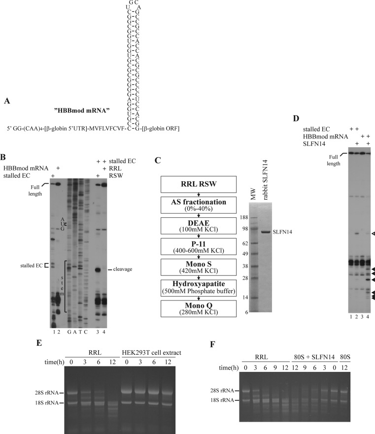
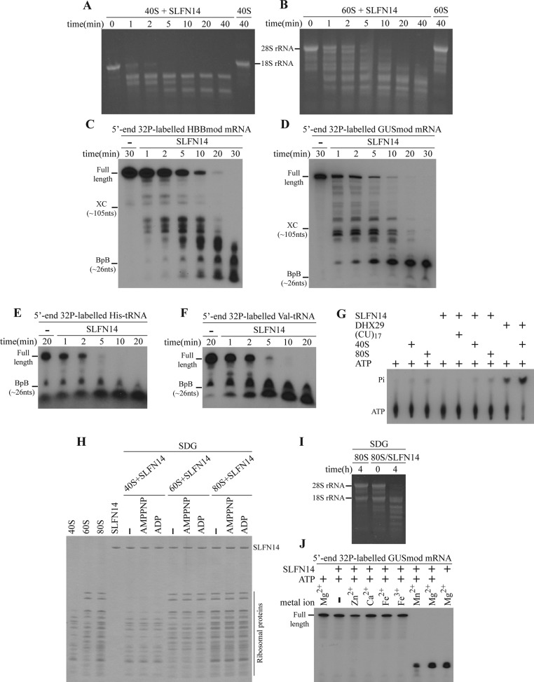
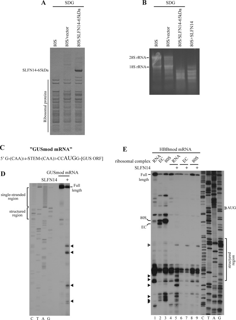
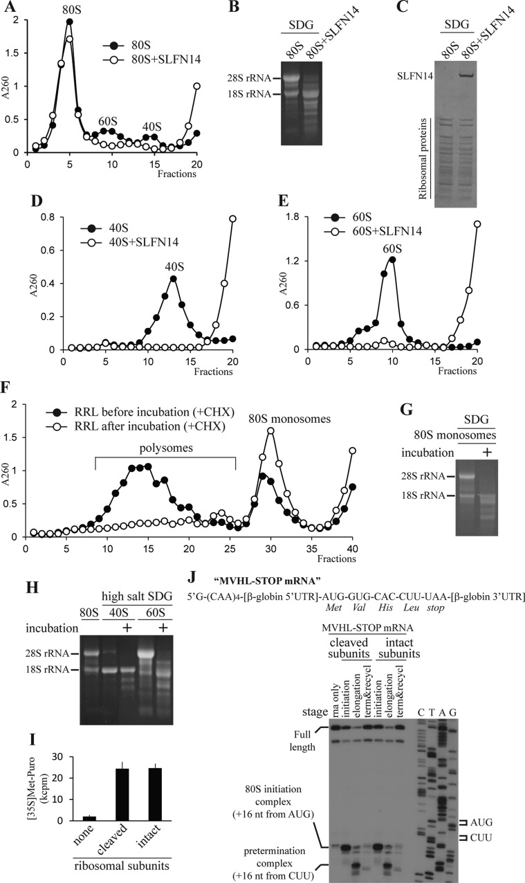
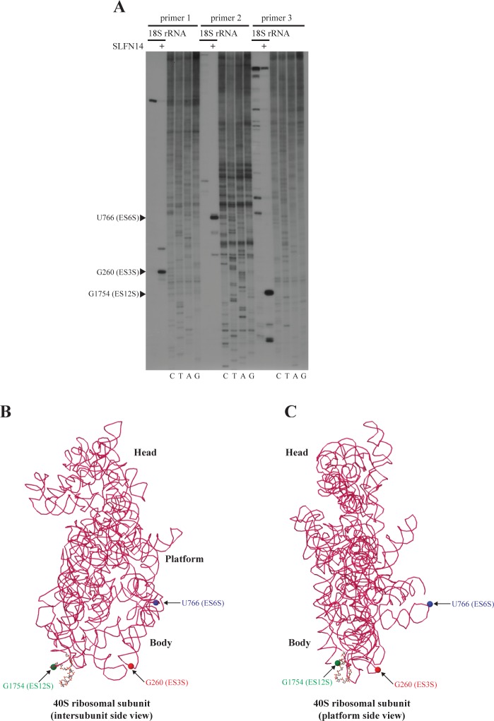
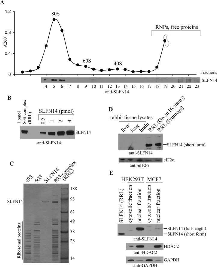
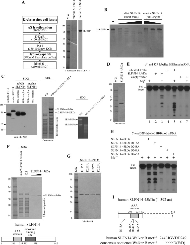
Turnover of mRNA is a critical step that allows cells to control gene expression. Endoribonucleases, enzymes cleaving RNA molecules internally, are some of the key components of the degradation process. Here we provide a detailed characterization of novel endoribonuclease SLFN14 purified from rabbit reticulocyte lysate. Schlafen genes encode a family of proteins limited to mammals. Their cellular function is unknown or incompletely understood. In reticulocytes, SLFN14 is strongly overexpressed, represented exclusively by the short form, all tethered to ribosomes, and appears to be one of the major ribosome-associated proteins. SLFN14 binds to ribosomes and ribosomal subunits in the low part of the body and cleaves RNA but preferentially rRNA and ribosome-associated mRNA. This results in the degradation of ribosomal subunits. This process is strictly Mg(2+)- and Mn(2+)-dependent, NTP-independent, and sequence nonspecific. However, in other cell types, SLFN14 is a full-length solely nuclear protein, which lacks ribosomal binding and nuclease activities. Mutational analysis revealed the ribosomal binding site and the aspartate essential for the endonucleolytic activity of protein. Only few endoribonucleases participating in ribosome-mediated processes have been characterized to date. Moreover, none of them are shown to be directly associated with the ribosome. Therefore, our findings expand the general knowledge of endoribonucleases involved in mammalian translation control.


Figures

Figure 1
Figure 1
Native rabbit C-terminally truncated SLFN14 causes endonucleolytic cleavage of rRNA in RRL and purified ribosomes as well as of mRNA in ribosome-associated and free states. (A) Structure of HBBmod mRNA. HBB is the name of the gene encoding β-globin protein. (B) Endonucleolytic cleavage of ribosome-bound HBBmod mRNA in stalled EC in the presence of RRL and RSW assayed by primer extension (lanes 3 and 4). Efficiency of stalled EC formation assembled from purified components in vitro analyzed by toeprint (lanes 1 and 2). Positions of full-length, stalled EC, and cleavage signals are indicated. Lanes G, A, T, and C depict the corresponding DNA sequence. (C) Purification scheme for SLFN14 (left). Purified SLFN14 resolved by SDS–PAGE (right). (D) Endonucleolytic cleavage of HBBmod mRNA in stalled EC and the free state by SLFN14 tested by primer extension. The empty triangle points to the cleavage position presented in both free and ribosome-associated mRNA. Black triangles indicate cleavage sites in free mRNA only. (E) Kinetics of rRNA degradation in RRL and HEK293T cell extract assayed by DAFGE. (F) Kinetics of degradation of rRNA by SLFN14 in RRL and purified 80S ribosomes analyzed by DAFGE.
Figure 2
Figure 2
SLFN14 binds the ribosome and ribosomal subunits and cleaves different types of RNA in a Mg2+- and Mn2+-dependent and ATP-independent manner. (A and B) Kinetics of degradation of rRNA by SLFN14 in purified 40S subunits and 60S subunits, respectively, assayed by DAFGE. (C–F) Kinetics of degradation of 5′-end 32P-labeled HBBmod mRNA, GUSmod mRNA, His-tRNA, and Val-tRNA, respectively, by SLFN14 analyzed by denaturing PAGE. (G) Thin layer chromatography analysis of SLFN14’s ATPase activity in the presence or absence of (CU)17 RNA, 40S subunits, or 80S ribosomes. DHX29 helicase (positive control) is ATPase; its hydrolyzing activity is stimulated by 40S subunits. Positions of [γ-32P]ATP and [32P]Pi are indicated. (H) Association of SLFN14 with 80S ribosomes or 40S and 60S subunits in the presence or absence of nucleotides, as indicated, assayed by SDG centrifugation and SDS–PAGE of peak ribosomal fractions. (I) rRNA degradation in the SDG-purified 80S/SLFN14 complex before or after incubation tested by DAFGE. (J) Dependence of SLFN14 endonucleolytic activity on metal ions and ATP analyzed by denaturing PAGE.
Figure 3
Figure 3
Endonucleolytic activity of recombinant human SLFN14–65kDa and sequence and structure specificity of SLFN14 in cleavage of free and ribosome-associated mRNA. (A) Affinity purification of SLFN14–65kDa on 80S ribosomes by centrifugation through SDG assayed by SDS–PAGE and Coomassie staining. (B) rRNA degradation in the SDG-purified 80S/SLFN14–65kDa complex, the 80S/empty vector expression complex, and the empty 80S ribosome after incubation analyzed by DAFGE. Native rabbit SLFN14 is a positive control (lane 4). (C) Structure of GUSmod mRNA. GUS is the name of the gene encoding β-glucuronidase protein. (D and E) Endonucleolytic cleavage of free GUSmod mRNA (D), free HBBmod mRNA (E, lanes 4 and 5), and ribosome-bound HBBmod mRNA (E, lanes 6–9) in 80S initiation (80S) and elongation complexes (EC) by SLFN14 assayed by primer extension. Efficiency of formation of ribosomal complexes assembled from purified components in vitro analyzed by toeprint (E, lanes 1–3). The empty triangle points to the cleavage position presented in both free and ribosome-associated mRNA. Black triangles indicate cleavage positions in free mRNA only. Positions of full-length cDNA and toeprints corresponding to ribosomal complexes as well as single-stranded and structured regions within mRNAs are indicated. Lanes C, T, A, and G depict corresponding DNA sequences.
Figure 4
Figure 4
Integrity and translation activity of ribosomes and ribosomal subunits after rRNA cleavage by SLFN14. (A) Integrity of 80S ribosomes after incubation with SLFN14 assayed by SDG centrifugation. (B and C) rRNA appearance and protein content, respectively, of 80S ribosomes after SLFN14 treatment and SDG purification analyzed by DAFGE and SDS–PAGE, respectively. (D and E) Degradation of 40S subunits and 60S subunits, respectively, after incubation with SLFN14 assayed by SDG centrifugation. (F) Ribosomal profiling of RRL before and after incubation at 37 °C in the presence of CHX obtained by SDG centrifugation. The total amount of ribosomes was decreased by 26 ± 3% (average ± standard deviation; n = 3) after incubation. Upper fractions were omitted for the sake of clarity. (G) rRNA appearance of 80S monosomes after incubation of RRL at 37 °C and ribosomal profiling. (H) rRNA appearance of cleaved 80S ribosome-derived 40S and 60S subunits purified from RRL after incubation at 37 °C and centrifugation through high-salt SDG. (I) Methionyl-puromycin assay of 80S initiation complex formation with purified intact/cleaved ribosomal subunits. (J) Structure of MVHL-STOP mRNA (top panel). Toeprint analysis of ribosomal complexes at different stages of translation assembled on MVHL-STOP mRNA in vitro with purified intact/cleaved ribosomal subunits (bottom). Lanes C, T, A, and G depict the cDNA sequence of MVHL-STOP mRNA. Positions of full-length cDNA and of toeprints corresponding to ribosomal complexes are indicated.
Figure 5
Figure 5
Ribosomal position of SLFN14. (A) Identification of prevalent cleavage sites in 18S rRNA after incubation of 40S ribosomal subunits with SLFN14 assayed by primer extension. Black triangles indicate the position and expansion segment location of residues cleaved by SLFN14. Primers 1–3 are described in the Supporting Information. (B and C) Intersubunit side and platform side views, respectively, of 18S rRNA nucleotides cleaved by SLFN14 mapped onto the crystal structure of the Saccharomyces cerevisiae 40S subunit (Protein Data Bank entry 3U5B). 18S rRNA (magenta) is shown in backbone representation. Protected nucleotides are colored green, red, and blue.
Figure 6
Figure 6
SLFN14 protein level and localization in different cell types. (A) SLFN14 distribution (bottom) in the ribosomal profile of mRNA-free RRL (Promega) (top) after SDG centrifugation assayed by immunoblotting. (B and C) Estimation of the SLFN14 to 80S ribosome ratio and SLFN14 abundance, respectively, in the SDG-purified 80S complex from RRL analyzed by immunoblotting and SDS–PAGE, respectively. (D) SLFN14 protein level in two different sources of RRL and rabbit brain, lung, and liver tissue lysates tested by immunoblotting with anti-SLFN14 and anti-eIF2α (loading control) antibodies. (E) SLFN14 subcellular distribution in HEK293T and MCF7 cells assayed by immunoblotting with anti-SLFN14, anti-HDAC2 (nuclear marker), and anti-GAPDH (cytoplasmic marker) antibodies.
Figure 7
Figure 7
Analysis of ribosomal binding and endonucleolytic activities of native murine full-length SLFN14 and mutational analysis of recombinant human SLFN14. (A) Purification scheme for native murine SLFN14 (left). Purified SLFN14 assayed by SDS–PAGE and immunoblotting (right). (B) Endonucleolytic cleavage of 18S rRNA in the 40S subunit by murine full-length and rabbit C-terminally truncated native forms of SLFN14 at different concentrations tested by denaturing PAGE. (C) Association of native murine and rabbit SLFN14 proteins with 40S subunits and 80S ribosomes assayed by SDG centrifugation and immunoblotting of peak ribosomal fractions (left). Association of recombinant murine SLFN14–65kDa with 80S ribosomes assayed by SDG centrifugation and Coomassie staining (middle). rRNA degradation in the SDG-purified 80S/murine SLFN14–65kDa complex and empty 80S ribosome after incubation analyzed by DAFGE (right). (D) Purified recombinant human SLFN14–45kDa resolved by SDS–PAGE. (E) Endonucleolytic activity of SLFN14–45kDa vs native rabbit SLFN14 analyzed by denaturing PAGE. The concentration of the recombinant or native form in the reaction is 0.9 μM or 11 nM, respectively. (F) Ribosomal binding of SLFN14–45kDa tested by SDG centrifugation and SDS–PAGE (top). Location of the ribosomal binding site in human SLFN14 (bottom). (G) Purified recombinant human SLFN14–45kDa mutants resolved by SDS–-PAGE. (H) Endonucleolytic activity of different SLFN14–45kDa point mutants assayed by denaturing PAGE. (I) Location of point mutations in SLFN14–45kDa.

References

    1. Li W. M.; Barnes T.; Lee C. H. (2010) Endoribonucleases: Enzymes gaining spotlight in mRNA metabolism. FEBS J. 277, 627–641. - PubMed
    1. Tomecki R.; Dziembowski A. (2010) Novel endoribonucleases as central players in various pathways of eukaryotic RNA metabolism. RNA 116, 1692–1724. - PMC - PubMed
    1. Walter P.; Ron D. (2011) The unfolded protein response: From stress pathway to homeostatic regulation. Science 334, 1081–1086. - PubMed
    1. Hollien J.; Weissman J. S. (2006) Decay of endoplasmic reticulum-localized mRNAs during the unfolded protein response. Science 313, 104–107. - PubMed
    1. Cunningham K. S.; Hanson M. N.; Schoenberg D. R. (2001) Polysomal ribonuclease 1 exists in a latent form on polysomes prior to estrogen activation of mRNA decay. Nucleic Acids Res. 29, 1156–1162. - PMC - PubMed

Publication types